Skip to main content Skip to navigation
Alexa Fluor® 647 Mouse Anti-Human CD307c
Multicolor flow cytometric analysis of human CD307c expression on human peripheral blood lymphocytes.          Left Panel:  Whole blood was stained with PerCP-Cy™5.5 Mouse Anti-Human CD19 antibody (Cat. No. 561295) and either Alexa Fluor® 647 Mouse IgG2b, κ Isotype Control (Cat. No. 558713; Left Plot) or Alexa Fluor® 647 Mouse Anti-Human CD307c antibody (Cat. No. 564768; Right Plot). Erythrocytes were lysed with BD PharmLyse™ Lysing Buffer (Cat. No. 555899). Two-color flow cytometric contour plots showing the correlated expression of CD307c (or Ig Isotype control staining) versus CD19 were derived from gated events with the forward and side light-scatter characteristics of viable lymphocytes. Right Panel: Human peripheral blood mononuclear cells were stained with FITC Mouse Anti-Human CD4 (Cat. No. 555346/561005/561842), PerCP-Cy™5.5 Mouse Anti-Human CD127 (Cat. No. 560551), BV421 Mouse Anti-Human CD25 (Cat. No. 562442/562443) antibodies, and either Alexa Fluor® 647 Mouse IgG2b, κ Isotype Control (Left Plot) or Alexa Fluor® 647 Mouse Anti-Human CD307c antibody (Right Plot). The cells were then fixed and permeabilized using the BD Pharmingen™ Transcription Factor Buffer Set (Cat. No. 562574/562725) and stained with PE Mouse Anti-Human FoxP3 antibody (Cat. No. 560046/560082). Flow cytometric contour plots showing the correlated expression of CD307c (or Ig Isotype control staining) versus FoxP3 were derived from gated events with the forward and side light-scatter characteristics of intact CD4+CD25+CD127low lymphocytes. Flow cytometric analysis was performed using a BD LSRFortessa™ Cell Analyzer System.
製品詳細
Down Arrow Up Arrow


BD Pharmingen™
FCRL3; Fc receptor-like 3; FcRH3; IRTA3; IFGP3; hIFGP3; SPAP2
Human (QC Testing)
Mouse IgG2b, κ
Recombinant Human FCRL3
Flow cytometry (Routinely Tested)
5 µl/test
IX 22
AB_2738940
Aqueous buffered solution containing BSA and ≤0.09% sodium azide.
RUO


Preparation and Storage

Store undiluted at 4°C and protected from prolonged exposure to light. Do not freeze. The monoclonal antibody was purified from tissue culture supernatant or ascites by affinity chromatography. The antibody was conjugated to Alexa Fluor® 647 under optimum conditions, and unreacted Alexa Fluor® 647 was removed.

Product Notices

  1. This reagent has been pre-diluted for use at the recommended Volume per Test. We typically use 1 × 10^6 cells in a 100-µl experimental sample (a test).
  2. An isotype control should be used at the same concentration as the antibody of interest.
  3. Alexa Fluor® 647 fluorochrome emission is collected at the same instrument settings as for allophycocyanin (APC).
  4. Odyssey™ Infrared Imaging System is available from LI-COR® Biosciences. For more information, contact 800 645 4267.
  5. Alexa Fluor® is a registered trademark of Molecular Probes, Inc., Eugene, OR.
  6. The Alexa Fluor®, Pacific Blue™, and Cascade Blue® dye antibody conjugates in this product are sold under license from Molecular Probes, Inc. for research use only, excluding use in combination with microarrays, or as analyte specific reagents. The Alexa Fluor® dyes (except for Alexa Fluor® 430), Pacific Blue™ dye, and Cascade Blue® dye are covered by pending and issued patents.
  7. Caution: Sodium azide yields highly toxic hydrazoic acid under acidic conditions. Dilute azide compounds in running water before discarding to avoid accumulation of potentially explosive deposits in plumbing.
  8. For fluorochrome spectra and suitable instrument settings, please refer to our Multicolor Flow Cytometry web page at www.bdbiosciences.com/colors.
  9. Please refer to www.bdbiosciences.com/us/s/resources for technical protocols.
  10. Source of all serum proteins is from USDA inspected abattoirs located in the United States.
564768 Rev. 1
抗体の詳細
Down Arrow Up Arrow
H5

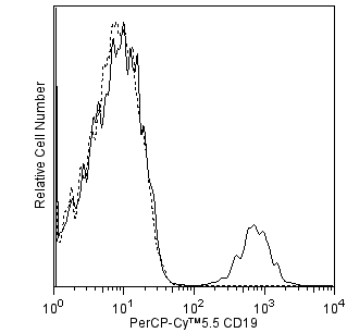
The H5 monoclonal antibody specifically binds to CD307c which is also known as Fc receptor-like 3 (FCRL3), Immune receptor translocation-associated protein 3 (IRTA3), IFGP family protein 3 (IFGP3), and SH2 domain-containing phosphatase anchor protein 2 (SPAP2). The FCRL3 gene is present in humans but not in mice. CD307c is a type I transmembrane glycoprotein that belongs to the FCRL family within the Ig gene superfamily. CD307c shares sequence homology with classical the Fc receptors. CD307c is expressed on subsets of B cells, plasma cells, NK cells, CD8+ T cells, and CD4+ natural T regulatory cells. CD307c isoforms contain multiple extracellular Ig domains and immunoreceptor-tyrosine activation (ITAM) and inhibitory (ITIM) motifs in their intracellular domains. CD307c may be involved in the regulation of immune responses. Genetic variation in FCRL3 has been associated with susceptibility to certain autoimmune diseases.

564768 Rev. 1
フォーマットの詳細
Down Arrow Up Arrow
Alexa Fluor™ 647
Alexa Fluor™ 647 Dye is part of the BD red family of dyes. This is a small organic fluorochrome with an excitation maximum (Ex Max) at 653-nm and an emission maximum (Em Max) at 669-nm. Alexa Fluor 647 is designed to be excited by the Red laser (627-640 nm) and detected using an optical filter centered near 520-nm (e.g., a 660/20 nm bandpass filter). Please ensure that your instrument’s configurations (lasers and optical filters) are appropriate for this dye.
Alexa Fluor™ 647
Red 627-640 nm
653 nm
669 nm
564768 Rev.1
引用&参考文献
Down Arrow Up Arrow

Development References (6)

  1. Kochi Y, Myouzen K, Yamada R, et al. FCRL3, an autoimmune susceptibility gene, has inhibitory potential on B-cell receptor-mediated signaling. J Immunol. 2009; 183(9):5502-5510. (Biology). View Reference
  2. Llinas L, Lazaro A, de Salort J, Matesanz-Isabel J, Sintes J, Engel P. Expression profiles of novel cell surface molecules on B-cell subsets and plasma cells as analyzed by flow cytometry. Immunol Lett. 2011; 134(2):113-121. (Clone-specific: Flow cytometry). View Reference
  3. Matesanz-Isabel J, Sintes J, Llinas L, de Salort J, Lazaro A, Engel P. New B-cell CD molecules. Immunol Lett. 2011; 134(2):104-112. (Clone-specific: Flow cytometry). View Reference
  4. Nagata S, Ise T, Pastan I. Fc receptor-like 3 protein expressed on IL-2 nonresponsive subset of human regulatory T cells. J Immunol. 2009; 182(12):7518-7526. (Immunogen: ELISA, Flow cytometry, Fluorescence activated cell sorting, Functional assay). View Reference
  5. Polson AG, Zheng B, Elkins K, et al. Expression pattern of the human FcRH/IRTA receptors in normal tissue and in B-chronic lymphocytic leukemia. Int Immunol. 2006; 18(9):1363-1373. (Biology). View Reference
  6. Swainson LA, Mold JE, Bajpai UD, McCune JM. Expression of the autoimmune susceptibility gene FcRL3 on human regulatory T cells is associated with dysfunction and high levels of programmed cell death-1. J Immunol. 2010; 184(7):3639-3647. (Biology). View Reference
すべて表示する (6) 表示項目を減らす
564768 Rev. 1

Please refer to Support Documents for Quality Certificates


Global - Refer to manufacturer's instructions for use and related User Manuals and Technical data sheets before using this products as described


Comparisons, where applicable, are made against older BD Technology, manual methods or are general performance claims.  Comparisons are not made against non-BD technologies, unless otherwise noted.

For Research Use Only. Not for use in diagnostic or therapeutic procedures.