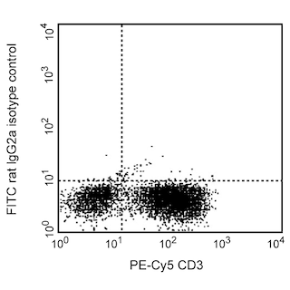
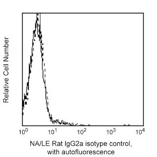
Skip to main content Skip to navigation
FITC Rat Anti-Human IL-2
Flow cytometric analysis of IL-2 expression by stimulated CD3+ human PBMC. Human PBMC were stimulated for 6 hours with PMA (Sigma, Cat. #P-8139) and calcium ionophore A23187 (Sigma, Cat. #C-9275) in the presence of 2 μM GolgiStop™ (Cat. No. 554724). The PBMC's were stained with PE-Cy™5 Mouse Anti-Human CD3 (Cat. No. 555334), fixed, permeabilized, and then stained with FITC Rat Anti-Human IL-2 (0.25 μg; Cat. No. 554565/561055/559361; left panel). To demonstrate specificity of staining, the binding of FITC-MQ1-17H12 was blocked by the preincubation of the conjugated antibody with a molar excess of recombinant human IL-2 protein (1.0 μg, Cat. No. 554603; middle panel), and by preincubation of the fixed/permeabilized cells with an excess of the Purified Rat Anti-Human IL-2 (10 μg, Cat. No. 554563; right panel) prior to staining with the FITC-MQ1-17H12 antibody. The quadrant markers for the bivariate dot plots were set based on the autofluorescence control, and verified with the blocking controls.
製品詳細
Down Arrow Up Arrow


BD Pharmingen™
IL2; Interleukin-2; T-cell growth factor; TCGF
Human (QC Testing), Rhesus, Cynomolgus, Baboon (Tested in Development)
Rat IgG2a, κ
Human IL-2 Recombinant Protein
Intracellular staining (flow cytometry) (Routinely Tested)
0.5 mg/ml
3558
AB_395482
Aqueous buffered solution containing ≤0.09% sodium azide.
RUO


Preparation and Storage

Store undiluted at 4°C and protected from prolonged exposure to light. Do not freeze. The monoclonal antibody was purified from tissue culture supernatant or ascites by affinity chromatography. The antibody was conjugated with FITC under optimum conditions, and unreacted FITC was removed.

推奨アッセイ手順

Immunofluorescent Staining and Flow Cytometry: The FITC-conjugated MQ1-17H12 antibody can be used for multicolor immunofluorescent staining and flow cytometric analysis to identify and enumerate IL-2-producing cells within mixed cell populations (see image). For optimal immunofluorescent staining for flow cytometric analysis, this anti-cytokine antibody should be titrated (≤ 0.5 µg mAb/1X10^6 cells)  For specific methodology, please visit the protocol section on our web site, http://www.bdbiosciences.com/resources/index.jsp

A useful control for demonstrating specificity of staining is either of the following: 1) pre-block the conjugated MQ1-17H12 antibody with its ligand (e.g., recombinant human IL-2; Cat. No. 554603) prior to staining, or 2) pre-block the fixed/permeabilized cells with unlabelled MQ1-17H12 antibody (Cat. No. 554563) prior to staining. The intracellular cytokine staining technique and use of blocking controls are described in detail by C. Prussin and D. Metcalfe. A suitable rat IgG2a isotype control for assessing the level of background staining on paraformaldehyde-fixed/saponin-permeabilized mouse or human cells is FITC-R35-95 (Cat. No. 554688); use at comparable concentrations to the antibody of interest (e.g., ≤ 0.5 µg mAb/1X10^6 cells).

Neutralization/Blocking: The NA/LE (Cat. No. 554562) format of the MQ1-17H12 antibody is useful for neutralization of human IL-2 bioactivity. A suitable NA/LE rat IgG2aisotype control is R35-95, Cat. No. 554687.

Product Notices

  1. Since applications vary, each investigator should titrate the reagent to obtain optimal results.
  2. An isotype control should be used at the same concentration as the antibody of interest.
  3. Caution: Sodium azide yields highly toxic hydrazoic acid under acidic conditions. Dilute azide compounds in running water before discarding to avoid accumulation of potentially explosive deposits in plumbing.
  4. For fluorochrome spectra and suitable instrument settings, please refer to our Multicolor Flow Cytometry web page at www.bdbiosciences.com/colors.
  5. Species cross-reactivity detected in product development may not have been confirmed on every format and/or application.
  6. Cy is a trademark of GE Healthcare.
  7. Please refer to www.bdbiosciences.com/us/s/resources for technical protocols.

関連製品

Stain Buffer (FBS) RUO
サイズ 500 mL カタログ番号 554656
sampleImage/
Stain Buffer (BSA) RUO
サイズ 500 mL カタログ番号 554657
sampleImage/
Protein Transport Inhibitor (Containing Monensin) RUO
サイズ 0.7 mL カタログ番号 554724
sampleImage/
FITC Rat IgG2a, κ Isotype Control RUO
サイズ 0.1 mg カタログ番号 554688
sampleImage/
Purified NA/LE Rat IgG2a, κ Isotype Control RUO
サイズ 0.5 mg カタログ番号 554687
sampleImage/
Recombinant Human IL-2 RUO
サイズ 10 µg カタログ番号 554603
sampleImage/
554565 Rev. 3
抗体の詳細
Down Arrow Up Arrow
MQ1-17H12

The MQ1-17H12 monoclonal antibody specifically binds to the multifunctional cytokine, human Interleukin-2 (IL-2). IL-2 is produced by activated T cells and has multiple functions that can affect the growth, proliferation, differentiation and survival of many different target cell types including T cells, B cells, NK cells, monocytes and macrophages. The immunogen used to generate the MQ1-17H12 hybridoma was purified recombinant human IL-2 protein. The MQ1-17H12 antibody reportedly neutralizes the biological activity of human IL-2.

554565 Rev. 3
フォーマットの詳細
Down Arrow Up Arrow
FITC
Fluorescein (FITC) is part of the BD blue family of dyes. This is a small organic fluorochrome with an excitation maximum (Ex Max) at 494-nm and an emission maximum (Em Max) at 518-nm. FITC is designed to be excited by the Blue laser (488-nm) and detected using an optical filter centered near 520 nm (e.g., a 530/30-nm bandpass filter). Please ensure that your instrument’s configurations (lasers and optical filters) are appropriate for this dye.
altImg
FITC
Blue 488 nm
494 nm
518 nm
554565 Rev.3
引用&参考文献
Down Arrow Up Arrow

Development References (3)

  1. Abrams J. Immunoenzymetric assay of mouse and human cytokines using NIP-labeled anti-cytokine antibodies. Curr Protoc Immunol. 2001; 1:6.20-6.21. (Biology). View Reference
  2. Abrams JS, Roncarolo MG, Yssel H, Andersson U, Gleich GJ, Silver JE. Strategies of anti-cytokine monoclonal antibody development: immunoassay of IL-10 and IL-5 in clinical samples. Immunol Rev. 1992; 127:5-24. (Biology). View Reference
  3. Prussin C, Metcalfe DD. Detection of intracytoplasmic cytokine using flow cytometry and directly conjugated anti-cytokine antibodies. J Immunol Methods. 1995; 188(1):117-128. (Clone-specific: Flow cytometry). View Reference
554565 Rev. 3

Please refer to Support Documents for Quality Certificates


Global - Refer to manufacturer's instructions for use and related User Manuals and Technical data sheets before using this products as described


Comparisons, where applicable, are made against older BD Technology, manual methods or are general performance claims.  Comparisons are not made against non-BD technologies, unless otherwise noted.

For Research Use Only. Not for use in diagnostic or therapeutic procedures.