
Preparation and Storage
Product Notices
- Since applications vary, each investigator should titrate the reagent to obtain optimal results.
- Please refer to www.bdbiosciences.com/us/s/resources for technical protocols.
- Caution: Sodium azide yields highly toxic hydrazoic acid under acidic conditions. Dilute azide compounds in running water before discarding to avoid accumulation of potentially explosive deposits in plumbing.
- Source of all serum proteins is from USDA inspected abattoirs located in the United States.
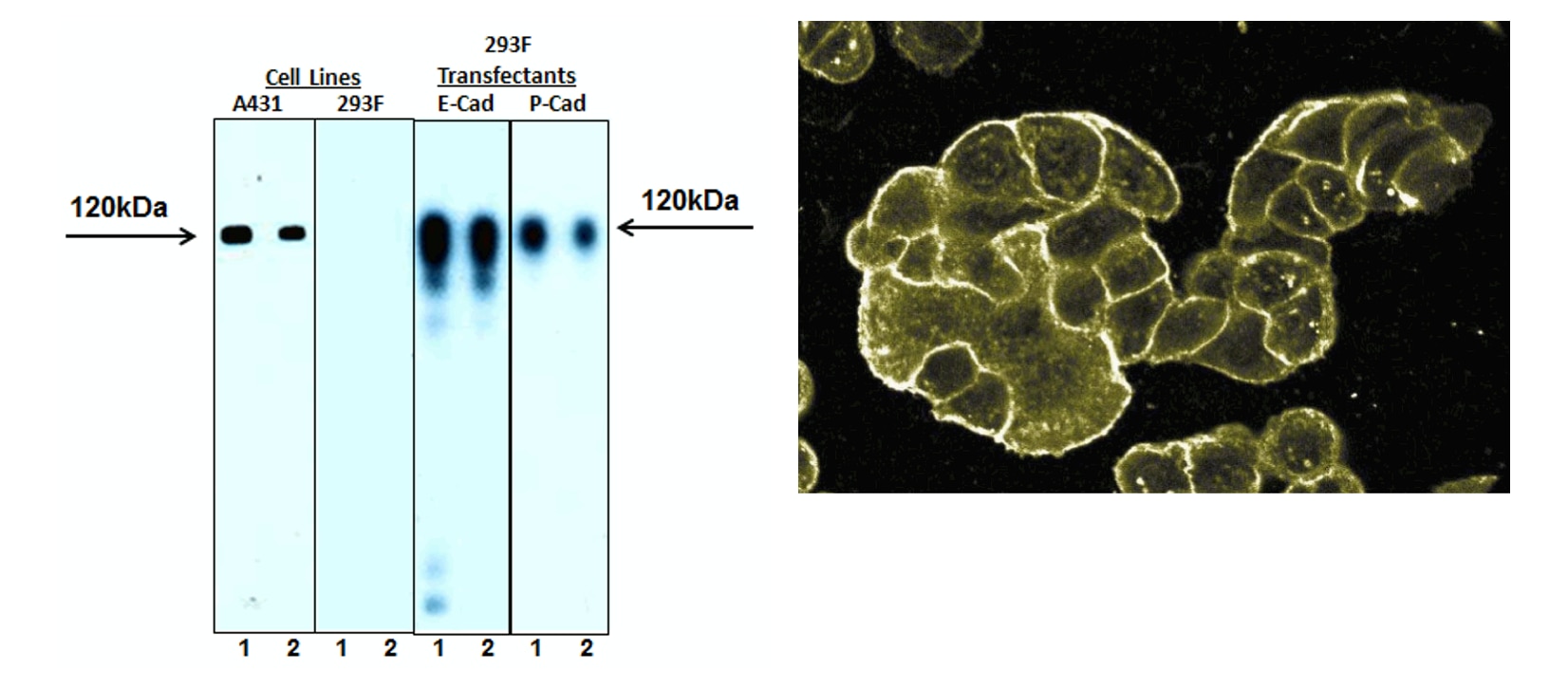
E-Cadherin is a 120-kDa transmembrane glycoprotein that is localized in the adherens junctions of epithelial cells. There it interacts with the cytoskeleton through the associated cytoplasmic catenin proteins. In addition to being a calcium-dependent adhesion molecule, E-Cadherin is also a critical regulator of epithelial junction formation. Its association with catenins is necessary for cell-cell adhesion. These E-cadherin/catenin complexes associate with cortical actin bundles at both the zonula adherens and the lateral adhesion plaques. Tyrosine phosphorylation can disrupt these complexes, leading to changes in cell adhesion properties. E-Cadherin expression is often down-regulated in highly invasive, poorly differentiated carcinomas. Increased expression of E-Cadherin in these cells reduces invasiveness. Thus, loss of expression or function of E-Cadherin appears to be an important step in tumorigenic progression. The 36/E-Cadherin monoclonal antibody recognizes the cytoplasmic domain of E-Cadherin, regardless of phosphorylation status. The peptide immunogen was generated from human E-Cadherin aa. 735-883.
Note: Investigators are advised that this antibody has some degree of cross-reactivity to P-Cadherin.
Development References (5)
-
Jaksits S, Kriehuber E, Charbonnier AS, Rappersberger K, Stingl G, Maurer D. CD34+ cell-derived CD14+ precursor cells develop into Langerhans cells in a TGF-beta 1-dependent manner. J Immunol. 1999; 163(9):4869-4877. (Clone-specific: Flow cytometry). View Reference
-
Miyoshi K, Shillingford JM, Smith GH, et al. Signal transducer and activator of transcription (Stat) 5 controls the proliferation and differentiation of mammary alveolar epithelium. J Cell Biol. 2001; 155(4):531-542. (Clone-specific: Immunohistochemistry). View Reference
-
Sheibani N, Sorenson CM, Frazier WA. Differential modulation of cadherin-mediated cell-cell adhesion by platelet endothelial cell adhesion molecule-1 isoforms through activation of extracellular regulated kinases. Mol Biol Cell. 2000; 11(8):2793-2802. (Clone-specific: Immunofluorescence, Western blot). View Reference
-
Takeichi M. The cadherins: cell-cell adhesion molecules controlling animal morphogenesis.. Development. 1988; 102(4):639-55. (Biology). View Reference
-
Weng Z, Xin M, Pablo L, et al. Protection against anoikis and down-regulation of cadherin expression by a regulatable beta-catenin protein. J Biol Chem. 2002; 277(21):18677-18686. (Clone-specific: Immunofluorescence, Immunoprecipitation, Western blot). View Reference
Please refer to Support Documents for Quality Certificates
Global - Refer to manufacturer's instructions for use and related User Manuals and Technical data sheets before using this products as described
Comparisons, where applicable, are made against older BD Technology, manual methods or are general performance claims. Comparisons are not made against non-BD technologies, unless otherwise noted.
For Research Use Only. Not for use in diagnostic or therapeutic procedures.