

Preparation and Storage
推奨アッセイ手順
Either BD Cytofix™ fixation buffer or BD Phosflow™ Fix Buffer I may be used for cell fixation. Any of the three BD Phosflow™ permeabilization buffers may be used.
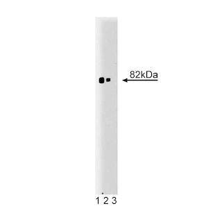
The purified format for this antibody has been reported to be reactive to human, mouse, rat, chicken, dog, and frog by western blotting (Cat. No. 610107 or 610108).
Product Notices
- This reagent has been pre-diluted for use at the recommended Volume per Test. We typically use 1 × 10^6 cells in a 100-µl experimental sample (a test).
- Alexa Fluor® is a registered trademark of Molecular Probes, Inc., Eugene, OR.
- Alexa Fluor® 647 fluorochrome emission is collected at the same instrument settings as for allophycocyanin (APC).
- The Alexa Fluor®, Pacific Blue™, and Cascade Blue® dye antibody conjugates in this product are sold under license from Molecular Probes, Inc. for research use only, excluding use in combination with microarrays, or as analyte specific reagents. The Alexa Fluor® dyes (except for Alexa Fluor® 430), Pacific Blue™ dye, and Cascade Blue® dye are covered by pending and issued patents.
- Source of all serum proteins is from USDA inspected abattoirs located in the United States.
- Caution: Sodium azide yields highly toxic hydrazoic acid under acidic conditions. Dilute azide compounds in running water before discarding to avoid accumulation of potentially explosive deposits in plumbing.
- For fluorochrome spectra and suitable instrument settings, please refer to our Multicolor Flow Cytometry web page at www.bdbiosciences.com/colors.
- Please refer to www.bdbiosciences.com/us/s/resources for technical protocols.
関連製品


The Protein Kinase C (PKC) family of homologous serine/threonine protein kinases is involved in a number of processes, such as growth, differentiation, and cytokine secretion. At least eleven isozymes have been described. These proteins are products of multiple genes and alternative splicing. Conventional PKC (cPKC) subfamily members (α, β, and γ isoforms) consists of a single polypeptide chain containing four conserved regions (C) and five variable regions (V). The N-terminal half containing C1, C2, V1, and V2 constitutes the regulatory domain and interacts with the PKC activators Ca2+, phospholipid, diacylglycerol, or phorbol ester. However, the the C2-like domains of novel PKC (nPKC) subfamily members (δ, ε, η, and θ isoforms) are Ca2+-independent. The atypical PKC (aPKC) subfamily members (ζ, ι, and λ isoforms) lack the C2 domain and are unique in that their activity is independent of diacylglycerols and phorbol esters. They also lack one repeat of the cysteine-rich sequences that are conserved in cPKC and nPKC members. The C-terminal region of PKC contains the catalytic domain. The PKC pathway represents a major signal transduction system that is activated following ligand-stimulation of transmembrane receptors by hormones, neurotransmitters, and growth factors. PKCα regulates a wide variety of functions such as cellular growth, apoptosis, cardiomyocyte function, and brain cognitive functions.
The 3/PKCα monoclonal antibody recognizes PKCα, regardless of phosphorylation status, and has been reported to crossreact with PKCβ.
Development References (6)
-
Bivona TG, Quatela SE, Bodemann BO, et al. PKC regulates a farnesyl-electrostatic switch on K-Ras that promotes its association with Bcl-Xl on mitochondria and induces apoptosis . Mol Cell. 2006; 21(4):481-493. (Biology). View Reference
-
Braz JC, Gregory K, Pathak A, et al. PKC-α regulates cardiac contractility and propensity toward heart failure. Nat Med. 2004; 10:248-254. (Biology). View Reference
-
Nishizuka Y. The molecular heterogeneity of protein kinase C and its implications for cellular regulation. Nature. 1988; 334(6184):661-665. (Biology). View Reference
-
Parker PJ, Murray-Rust J. PKC at a glance. J Cell Sci. 2004; 117:131-132. (Biology). View Reference
-
Zhang XA, Bontrager AL, Hemler ME.. Transmembrane-4 superfamily proteins associate with activated protein kinase C (PKC) and link PKC to specific beta(1) integrins. J Biol Chem. 2001; 276(27):25005-25013. (Clone-specific: Immunofluorescence, Immunoprecipitation, Western blot). View Reference
-
de Quervain D J-F, Papassotiropoulos A. Identification of a genetic cluster influencing memory performance and hippocampal activity in humans. Proc Natl Acad Sci U S A. 2006; 103(11):4270-4274. (Biology). View Reference
Please refer to Support Documents for Quality Certificates
Global - Refer to manufacturer's instructions for use and related User Manuals and Technical data sheets before using this products as described
Comparisons, where applicable, are made against older BD Technology, manual methods or are general performance claims. Comparisons are not made against non-BD technologies, unless otherwise noted.
For Research Use Only. Not for use in diagnostic or therapeutic procedures.