Skip to main content Skip to navigation
Alexa Fluor® 647 Mouse Anti-Sox2
Flow cytometric analysis of Sox2 on human embryonic stem (ES) cells. H7 human ES cells (WiCell, Madison, WI) passage 46 grown on irradiated mouse embryonic fibroblasts were harvested with Accutase™ (Cat No. 561527) and fixed with BD Cytofix™ Fixation Buffer (Cat. No. 554655). Cells were permeablized with BD™ Phosflow Perm Buffer III (Cat No. 558050) and stained with Alexa Fluor® 647 Mouse anti-Human Sox2 antibody (solid line) or Alexa Fluor® 647 mouse IgG1, κ isotype control (Clone MOPC-21, Cat. No. 557714, dashed line). Flow cytometry was performed on a BD LSR™ II flow cytometry system.
Alexa Fluor® 647 Mouse Anti-Sox2
Immunoflourescent staining of Sox2 on human embryonic stem (ES) cells. H9 human ES cells (WiCell, Madison, WI) passage 39 grown in mTeSR™1 medium (StemCell Technologies) on BD Matrigel™ hESC-qualified Matrix (Cat. No. 354277) were fixed with BD Cytofix™ Fixation Buffer (Cat. No. 554655). Cells were permeablized with BD™ Phosflow Perm Buffer III (Cat No. 558050) and stained with Alexa Fluor® 647 Mouse anti-Sox2 monoclonal antibody (pseudo colored red) at 5 µg/mL. Counter-staining of cell nuclei was with Hoechst 33342 (pseudo-colored blue). The images were captured on a BD Pathway™ 435 Cell Analyzer and merged using BD Attovision™ Software.
Flow cytometric analysis of Sox2 on human embryonic stem (ES) cells. H7 human ES cells (WiCell, Madison, WI) passage 46 grown on irradiated mouse embryonic fibroblasts were harvested with Accutase™ (Cat No. 561527) and fixed with BD Cytofix™ Fixation Buffer (Cat. No. 554655). Cells were permeablized with BD™ Phosflow Perm Buffer III (Cat No. 558050) and stained with Alexa Fluor® 647 Mouse anti-Human Sox2 antibody (solid line) or Alexa Fluor® 647 mouse IgG1, κ isotype control (Clone MOPC-21, Cat. No. 557714, dashed line). Flow cytometry was performed on a BD LSR™ II flow cytometry system.
Immunoflourescent staining of Sox2 on human embryonic stem (ES) cells. H9 human ES cells (WiCell, Madison, WI) passage 39 grown in mTeSR™1 medium (StemCell Technologies) on BD Matrigel™ hESC-qualified Matrix (Cat. No. 354277) were fixed with BD Cytofix™ Fixation Buffer (Cat. No. 554655). Cells were permeablized with BD™ Phosflow Perm Buffer III (Cat No. 558050) and stained with Alexa Fluor® 647 Mouse anti-Sox2 monoclonal antibody (pseudo colored red) at 5 µg/mL. Counter-staining of cell nuclei was with Hoechst 33342 (pseudo-colored blue). The images were captured on a BD Pathway™ 435 Cell Analyzer and merged using BD Attovision™ Software.
製品詳細
Down Arrow Up Arrow


BD Pharmingen™
Human (QC Testing), Mouse (Reactivity Confirmed in Development)
Mouse CD, also known as Charles River SD (outbred) IgG1, κ
Human Sox2 Recombinant Protein
Intracellular staining (flow cytometry) (Routinely Tested), Bioimaging, Immunofluorescence (Tested During Development)
5 µl
AB_10897844
Aqueous buffered solution containing BSA and ≤0.09% sodium azide.
RUO


Preparation and Storage

Store undiluted at 4°C and protected from prolonged exposure to light. Do not freeze. The monoclonal antibody was purified from tissue culture supernatant or ascites by affinity chromatography. The antibody was conjugated to Alexa Fluor® 647 under optimum conditions, and unreacted Alexa Fluor® 647 was removed.

Product Notices

  1. This reagent has been pre-diluted for use at the recommended Volume per Test. We typically use 1 × 10^6 cells in a 100-µl experimental sample (a test).
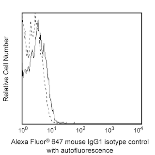
  2. An isotype control should be used at the same concentration as the antibody of interest.
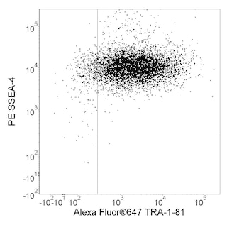
  3. Alexa Fluor® 647 fluorochrome emission is collected at the same instrument settings as for allophycocyanin (APC).
  4. Alexa Fluor® is a registered trademark of Molecular Probes, Inc., Eugene, OR.
  5. Source of all serum proteins is from USDA inspected abattoirs located in the United States.
  6. Caution: Sodium azide yields highly toxic hydrazoic acid under acidic conditions. Dilute azide compounds in running water before discarding to avoid accumulation of potentially explosive deposits in plumbing.
  7. For fluorochrome spectra and suitable instrument settings, please refer to our Multicolor Flow Cytometry web page at www.bdbiosciences.com/colors.
  8. Please refer to www.bdbiosciences.com/us/s/resources for technical protocols.
  9. The Alexa Fluor®, Pacific Blue™, and Cascade Blue® dye antibody conjugates in this product are sold under license from Molecular Probes, Inc. for research use only, excluding use in combination with microarrays, or as analyte specific reagents. The Alexa Fluor® dyes (except for Alexa Fluor® 430), Pacific Blue™ dye, and Cascade Blue® dye are covered by pending and issued patents.
  10. mTESR™1 is a trademark of StemCell Technologies.
562139 Rev. 1
抗体の詳細
Down Arrow Up Arrow
O30-678

The monoclonal antibody O30-678 recognizes the Sox2 transcription factor. Sox2  [SRY (sex determining region Y)-box 2] is a member of the  SRY-related HMG-box (SOX) family of transcription factors. Sox2 is required for the maintenance of the undifferentiated state of pluripotent stem cells. Complexes of Sox2 with the homeobox transcription factors Oct3/4 and/or Nanog bind to the promoters of a network of genes that are involved in the maintenance of pluripotency and self renewal in stem cells. Sox2 is also a marker of neural stem cells during embryonic development and in the adult brain. The O30-678 antibody recognizes both human and mouse Sox2 proteins.  

562139 Rev. 1
フォーマットの詳細
Down Arrow Up Arrow
Alexa Fluor™ 647
Alexa Fluor™ 647 Dye is part of the BD red family of dyes. This is a small organic fluorochrome with an excitation maximum (Ex Max) at 653-nm and an emission maximum (Em Max) at 669-nm. Alexa Fluor 647 is designed to be excited by the Red laser (627-640 nm) and detected using an optical filter centered near 520-nm (e.g., a 660/20 nm bandpass filter). Please ensure that your instrument’s configurations (lasers and optical filters) are appropriate for this dye.
Alexa Fluor™ 647
Red 627-640 nm
653 nm
669 nm
562139 Rev.1
引用&参考文献
Down Arrow Up Arrow

Development References (3)

  1. Boyer LA, Lee TI, Cole MF, et al. Core transcriptional regulatory circuitry in human embryonic stem cells. Cell. 2005; 122:947-956. (Biology). View Reference
  2. Pan G, Thomson JA. Nanog and transcriptional networks in embryonic stem cell pluripotency. Cell Res. 2007; 17:42-49. (Biology). View Reference
  3. Takahashi K, Yamanaka S. Induction of pluripotent stem cells from mouse embryonic and adult fibroblast cultures by defined factors. Cell. 2006; 126(4):663-676. (Biology). View Reference
562139 Rev. 1

Please refer to Support Documents for Quality Certificates


Global - Refer to manufacturer's instructions for use and related User Manuals and Technical data sheets before using this products as described


Comparisons, where applicable, are made against older BD Technology, manual methods or are general performance claims.  Comparisons are not made against non-BD technologies, unless otherwise noted.

For Research Use Only. Not for use in diagnostic or therapeutic procedures.