Skip to main content Skip to navigation
Alexa Fluor® 647 Mouse anti-IκBα
Flow cytometric analysis of IκBα expression. LEFT: IκBα expression in HeLa cells. HeLa cells (ATCC CCL-2) were either treated (shaded histogram) with 20 ng/mL recombinant human TNF (Cat. No. 554618) for 10 min at 37°C or untreated (open histogram). The cells were fixed (BD Cytofix™ Fixation Buffer, Cat. No. 554655) for 10 minutes at 37°C, then permeabilized (BD Phosflow™ Perm Buffer II, Cat. No. 558052) on ice for at least 30 minutes, and then stained with Alexa Fluor® 647 Mouse anti-IκBα antibody. The fluorescence histograms were derived from gated events with the forward and side light-scatter characteristics of intact cells. RIGHT: IκBα expression in human peripheral blood T cells. Human whole blood (collected with heparin) was either unstimulated (open histogram) or stimulated (shaded histogram) with 400 nM PMA plus 250 ng/mL ionomycin (Sigma, Cat. No. I-0634) for 15 min at 37°C. The erythrocytes were lysed and the leukocytes were fixed with BD Phosflow™ Lyse/Fix Buffer (Cat. No. 558049). The leukocytes were permeabilized with BD Phosflow™ Perm Buffer II (558052) on ice for at least 30 minutes, and then stained with Alexa Fluor® 647 Mouse anti-IκBα and BD Horizon™ V450 Mouse Anti-Human CD3 (Cat. No. 560365) antibodies. The fluorescence histograms were derived from human CD3-positive T cell-gated events with the forward and side light-scatter characteristics of intact lymphocytes. Flow cytometry was performed using a BD FACSCanto™ II flow cytometry system.
製品詳細
Down Arrow Up Arrow


BD Phosflow™
MAD-3; I-kappa-B-alpha; IkB-alpha; NFKBIA; NF-kappa-B inhibitor alpha
Human (QC Testing)
Mouse IgG1
Human IκBα aa. 145-302 Recombinant Protein
Intracellular staining (flow cytometry) (Routinely Tested)
5 µl
AB_2033974
Aqueous buffered solution containing BSA and ≤0.09% sodium azide.
RUO


Preparation and Storage

The monoclonal antibody was purified from tissue culture supernatant or ascites by affinity chromatography. The antibody was conjugated to Alexa Fluor® 647 under optimum conditions, and unreacted Alexa Fluor® 647 was removed. Store undiluted at 4°C and protected from prolonged exposure to light. Do not freeze.

推奨アッセイ手順

This Alexa Fluor® 647-conjugated antibody is suitable for intracellular staining of cell lines and primary cells using BD Cytofix™ Fixation Buffer or BD Phosflow™ Lyse/Fix Buffer. Although this antibody can be used with BD Phosflow™ Perm/Wash Buffer I or BD Phosflow™ Perm Buffers II, III or IV, it performs optimally when used with BD Phosflow™ Perm Buffer II.

Product Notices

  1. This reagent has been pre-diluted for use at the recommended Volume per Test. We typically use 1 × 10^6 cells in a 100-µl experimental sample (a test).
  2. Please refer to www.bdbiosciences.com/us/s/resources for technical protocols.
  3. The Alexa Fluor®, Pacific Blue™, and Cascade Blue® dye antibody conjugates in this product are sold under license from Molecular Probes, Inc. for research use only, excluding use in combination with microarrays, or as analyte specific reagents. The Alexa Fluor® dyes (except for Alexa Fluor® 430), Pacific Blue™ dye, and Cascade Blue® dye are covered by pending and issued patents.
  4. Alexa Fluor® is a registered trademark of Molecular Probes, Inc., Eugene, OR.
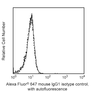
  5. Alexa Fluor® 647 fluorochrome emission is collected at the same instrument settings as for allophycocyanin (APC).
  6. For fluorochrome spectra and suitable instrument settings, please refer to our Multicolor Flow Cytometry web page at www.bdbiosciences.com/colors.
  7. Caution: Sodium azide yields highly toxic hydrazoic acid under acidic conditions. Dilute azide compounds in running water before discarding to avoid accumulation of potentially explosive deposits in plumbing.
  8. Source of all serum proteins is from USDA inspected abattoirs located in the United States.
  9. An isotype control should be used at the same concentration as the antibody of interest.
560817 Rev. 2
抗体の詳細
Down Arrow Up Arrow
25/IkBa/MAD-3

NF-κB is a transcription factor that is a member of the mammalian NF-κB/Rel family of proteins. Members of this family are involved in the regulation of cell proliferation, immune function, as well as development. In resting cells, IκBα binds to and maintains NF-κB in the cytoplasm by blocking the nuclear localization sequences of NF-κB. In the cellular response to an extracellular signal, IκBα is phosphorylated and subsequently degraded via the ubiquination-proteasome pathway, allowing NF-κB to translocate to the nucleus. Once in the nucleus, NF-κB can induce the transcription of IκBα thereby renewing the cycle so that IκBα can form a complex with NF-κB and maintain it in its cytoplasmic location. IκBα -/- mice show an increased level of NF-κB activity and have been shown to die soon after birth.

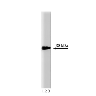
The 25/IkBa/MAD-3 monoclonal antibody recognizes human IκBα regardless of phosphorylation status and does not cross-react with mouse IκBα.

560817 Rev. 2
フォーマットの詳細
Down Arrow Up Arrow
Alexa Fluor™ 647
Alexa Fluor™ 647 Dye is part of the BD red family of dyes. This is a small organic fluorochrome with an excitation maximum (Ex Max) at 653-nm and an emission maximum (Em Max) at 669-nm. Alexa Fluor 647 is designed to be excited by the Red laser (627-640 nm) and detected using an optical filter centered near 520-nm (e.g., a 660/20 nm bandpass filter). Please ensure that your instrument’s configurations (lasers and optical filters) are appropriate for this dye.
Alexa Fluor™ 647
Red 627-640 nm
653 nm
669 nm
560817 Rev.2
引用&参考文献
Down Arrow Up Arrow

Development References (5)

  1. Auphan N, DiDonato JA, Rosette C, Helmberg A, Karin M. Immunosuppression by glucocorticoids: inhibition of NF-kappa B activity through induction of I kappa B synthesis. Science. 1995; 270(5234):286-290. (Biology). View Reference
  2. Cordle SR, Donald R, Read MA, Hawiger J. Lipopolysaccharide induces phosphorylation of MAD3 and activation of c-Rel and related NF-kappa B proteins in human monocytic THP-1 cells. J Biol Chem. 1993; 268(16):11803-11810. (Biology). View Reference
  3. Haskill S, Beg AA, Tompkins SM, et al. Characterization of an immediate-early gene induced in adherent monocytes that encodes I kappa B-like activity. Cell. 1991; 65(7):1281-1289. (Biology). View Reference
  4. Nakashio A, Fujita N, Rokudai S, Sato S, Tsuruo T. Prevention of phosphatidylinositol 3'-kinase-Akt survival signaling pathway during topotecan-induced apoptosis. Cancer Res. 2000; 60(18):5303-5309. (Clone-specific: Western blot). View Reference
  5. Traenckner EB, Pahl HL, Henkel T, Schmidt KN, Wilk S, Baeuerle PA. Phosphorylation of human I kappa B-alpha on serines 32 and 36 controls I kappa B-alpha proteolysis and NF-kappa B activation in response to diverse stimuli. EMBO J. 1995; 14(12):2876-2883. (Biology). View Reference
すべて表示する (5) 表示項目を減らす
560817 Rev. 2

Please refer to Support Documents for Quality Certificates


Global - Refer to manufacturer's instructions for use and related User Manuals and Technical data sheets before using this products as described


Comparisons, where applicable, are made against older BD Technology, manual methods or are general performance claims.  Comparisons are not made against non-BD technologies, unless otherwise noted.

For Research Use Only. Not for use in diagnostic or therapeutic procedures.