BB515 Mouse Anti-Human CD23
Clone M-L233 (RUO)
- Brand BD Horizon™
- Alternative Name FCER2; FcεRII; Low affinity immunoglobulin epsilon Fc receptor; BLAST-2
- Vol. Per Test 5 µl
- Isotype Mouse IgG1, κ
- Reactivity Human (QC Testing)
- Application
Flow cytometry (Routinely Tested)
- Workshop No. V CD23.15
- Storage Buffer Aqueous buffered solution containing ≤0.09% sodium azide.
- Regulatory Status RUO
Regulatory Status Legend
Description
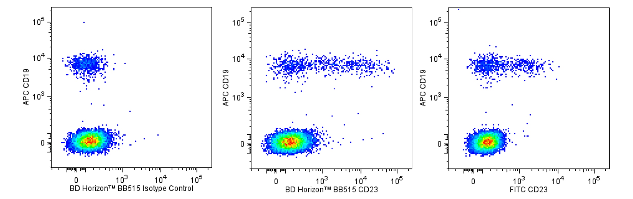
The M-L233 antibody specifically binds to human CD23, the low affinity receptor for human IgE (FcεRII). CD23 is a type II membrane glycoprotein that is expressed by B cells, monocytes, macrophages, eosinophils, platelets and dendritic cells. CD23 mediates IgE-dependent cytotoxicity and phagocytosis by macrophages and eosinophils. Soluble CD23 (sCD23) can be released by CD23-positive cells as a result of proteolytic cleavage of membrane CD23. Larger fragments of sCD23 (e.g., 25-37 kDa) retain their IgE-binding capacity whereas smaller fragments (ie, ≤ 12 kDa) do not. Soluble CD23 may have immunoregulatory effects on the growth and differentiation of B cells and other cell types.
The antibody was conjugated to BD Horizon BB515 which was developed exclusively by BD Biosciences. With an excitation max of 490 nm and an emission max of 515 nm, BD Horizon BB515 can be excited by the 488 nm laser and detected in a standard FITC set (eg, 530/30-nm filter). This dye provides a much brighter alternative to FITC with less spillover into the PE detector.
Format
BB515 is a dye that was exclusively developed by BD Biosciences as a brighter alternative to FITC. This dye is up to seven times brighter than FITC and has less spillover into the PE channel. Due to similar excitation and emission properties, BB515, FITC, and Alexa Fluor® 488 cannot be used simultaneously.
Suggested Companion Products
Stain Buffer (FBS) RUO
500 mL
Cat No: 554656
Stain Buffer (BSA) RUO
500 mL
Cat No: 554657
Brilliant Stain Buffer RUO
100 Tests
Cat No: 563794
BB515 Mouse IgG1, κ Isotype Control RUO
100 µg
Cat No: 564416
APC Mouse Anti-Human CD19 HIB19 RUO
100 Tests
Cat No: 555415
APC Mouse Anti-Human CD19 HIB19 RUO
25 Tests
Cat No: 561742
Lysing Solution 10X Concentrate RUO (GMP)
100
Cat No: 349202
Lysing Buffer RUO
100 mL
Cat No: 555899
Resources & Tools | ||||||
---|---|---|---|---|---|---|
Spectrum Viewer | Panel Designer | Spectrum Viewer | Download TDS | Regulatory Document Website |
Preparation and Storage
Store undiluted at 4°C and protected from prolonged exposure to light. Do not freeze.
The monoclonal antibody was purified from tissue culture supernatant or ascites by affinity chromatography.
The antibody was conjugated with BD Horizon™ BB515 under optimum conditions and unconjugated antibody was removed.
Product Notices
- This reagent has been pre-diluted for use at the recommended Volume per Test. We typically use 1 × 10^6 cells in a 100-µl experimental sample (a test).
- An isotype control should be used at the same concentration as the antibody of interest.
- Caution: Sodium azide yields highly toxic hydrazoic acid under acidic conditions. Dilute azide compounds in running water before discarding to avoid accumulation of potentially explosive deposits in plumbing.
- The manufacture, use, sale, offer for sale, or import of this product is subject to one or more patents or pending applications. This product, and only in the amount purchased by buyer, may be used solely for buyer’s own internal research, in a manner consistent with the accompanying product literature. No other right to use, sell or otherwise transfer (a) this product, or (b) its components is hereby granted expressly, by implication or by estoppel. Diagnostic uses require a separate license.
- For fluorochrome spectra and suitable instrument settings, please refer to our Multicolor Flow Cytometry web page at www.bdbiosciences.com/colors.
- Please refer to www.bdbiosciences.com/pharmingen/protocols for technical protocols.
For optimal results, it is recommended to perform 2 washes after staining with antibodies. Cells may be prepared, stained with antibodies and washed twice with wash buffer per established protocols for immunofluorescent staining, prior to acquisition on a flow cytometer. Performing fewer than the recommended wash steps may lead to increased spread of the negative population.