

Preparation and Storage
推奨アッセイ手順
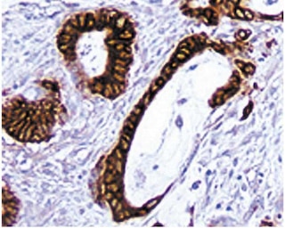
Immunohistochemistry: The HMPV clone reactive against human MUC1 is recommended to test for immunohistochemical staining of formalin-fixed paraffin and acetone-fixed frozen sections. For paraffin sections, microwave pretreatment with BD Retrievagen A (pH 6.5) (Cat. No. 550524) is required. Tissue tested was human breast cancer tissue. The antibody stains normal and cancerous cells with a high carbohydrate content. The isotype control recommended for use with this antibody is purified mouse IgG1 (Cat. No. 550878). For optimal indirect immunohistochemical staining, HMPV antibody should be titrated (1:10 to 1:50 dilution) and visualized via a three-step staining procedure in combination with polyclonal, biotin conjugated anti-mouse Igs (multiple adsorbed) (Cat. No. 550337) as the secondary antibody and Streptavidin-HRP (Cat. No. 550946) together with the DAB detection system (Cat. No. 550880). More conveniently, the Anti-Mouse Ig HRP detection kit (Cat. No. 551011) that contains the biotinylated secondary antibody, antibody diluent, Streptavidin-HRP and DAB substrate can be used for staining. A detailed protocol of the immunohistochemical procedure is available at our website, http://www.bdbiosciences.com/support/resources.
Product Notices
- Since applications vary, each investigator should titrate the reagent to obtain optimal results.
- Caution: Sodium azide yields highly toxic hydrazoic acid under acidic conditions. Dilute azide compounds in running water before discarding to avoid accumulation of potentially explosive deposits in plumbing.
- Source of all serum proteins is from USDA inspected abattoirs located in the United States.
- An isotype control should be used at the same concentration as the antibody of interest.
- This antibody has been developed for the immunohistochemistry application. However, a routine immunohistochemistry test is not performed on every lot. Researchers are encouraged to titrate the reagent for optimal performance.
- Sodium azide is a reversible inhibitor of oxidative metabolism; therefore, antibody preparations containing this preservative agent must not be used in cell cultures nor injected into animals. Sodium azide may be removed by washing stained cells or plate-bound antibody or dialyzing soluble antibody in sodium azide-free buffer. Since endotoxin may also affect the results of functional studies, we recommend the NA/LE (No Azide/Low Endotoxin) antibody format, if available, for in vitro and in vivo use.
- Please refer to www.bdbiosciences.com/us/s/resources for technical protocols.
関連製品


The HMPV monoclonal antibody specifically binds to CD227 which is also known as Mucin-1 (MUC1). A major form of CD227 is expressed as a type I transmembrane glycoprotein. CD227 belongs to the epithelial mucin family whose members are heavily O-glycosylated and characterized by high molecular weight, and an amino acid composition rich in serine, threonine, proline, and glycine. CD227 is variably expressed on the surfaces of normal and malignant glandular and ductal epithelial cells, and some hematopoietic cell lineages including subsets of T cells, B cells, monocytes and dendritic cells. Soluble forms of CD227 may arise by shedding from the cell surface or by secretion of forms derived from alternative RNA splicing. The HMPV antibody binds to the core peptide of the MUC1 protein. The core protein contains a domain of 20 amino-acid tandem repeats which function as multiple epitopes for this monoclonal antibody. Incomplete glycosylation of some tumor-associated mucins may lead to variable unmasking of the multiple peptide epitopes leading to the observed differences in immunostaining intensities between cells from normal and malignant tissues. CD227 plays roles in the provision of protective barrier function, the regulation of cellular adhesion, and the transduction of multiple signal pathways.
Development References (3)
-
Devine PL, Birrell GW, Whitehead RH, Harada H, Xing PX, McKenzie IF. Expression of MUC1 and MUC2 mucins by human tumor cell lines. Tumour Biol. 1992; 13(5):268-277. (Biology). View Reference
-
Xing PX, Prenzoska J, Layton GT, Devine PL, McKenzie IF. Second-generation monoclonal antibodies to intestinal MUC2 peptide reactive with colon cancer. J Natl Cancer Inst. 1992; 84(9):699-703. (Biology). View Reference
-
Xing PX, Prenzoska J, Quelch K, McKenzie IF. Second generation anti-MUC1 peptide monoclonal antibodies. Cancer Res. 1992; 52(8):2310-2317. (Biology). View Reference
Please refer to Support Documents for Quality Certificates
Global - Refer to manufacturer's instructions for use and related User Manuals and Technical data sheets before using this products as described
Comparisons, where applicable, are made against older BD Technology, manual methods or are general performance claims. Comparisons are not made against non-BD technologies, unless otherwise noted.
For Research Use Only. Not for use in diagnostic or therapeutic procedures.