-
抗体試薬
- フローサイトメトリー用試薬
-
ウェスタンブロッティング抗体試薬
- イムノアッセイ試薬
-
シングルセル試薬
- BD® AbSeq Assay | シングルセル試薬
- BD Rhapsody™ Accessory Kits | シングルセル試薬
- BD® Single-Cell Multiplexing Kit | シングルセル試薬
- BD Rhapsody™ Targeted mRNA Kits | シングルセル試薬
- BD Rhapsody™ Whole Transcriptome Analysis (WTA) Amplification Kit | シングルセル試薬
- BD Rhapsody™ TCR/BCR Profiling Assays (VDJ Assays) | シングルセル試薬
- BD® OMICS-Guard Sample Preservation Buffer
- BD Rhapsody™ ATAC-Seq Assays
-
細胞機能評価のための試薬
-
顕微鏡・イメージング用試薬
-
細胞調製・分離試薬
-
- BD® AbSeq Assay | シングルセル試薬
- BD Rhapsody™ Accessory Kits | シングルセル試薬
- BD® Single-Cell Multiplexing Kit | シングルセル試薬
- BD Rhapsody™ Targeted mRNA Kits | シングルセル試薬
- BD Rhapsody™ Whole Transcriptome Analysis (WTA) Amplification Kit | シングルセル試薬
- BD Rhapsody™ TCR/BCR Profiling Assays (VDJ Assays) | シングルセル試薬
- BD® OMICS-Guard Sample Preservation Buffer
- BD Rhapsody™ ATAC-Seq Assays
- Japan (Japanese)
-
Change country/language
Old Browser
Looks like you're visiting us from {countryName}.
Would you like to stay on the current country site or be switched to your country?


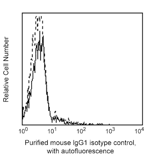
Multiparameter flow cytometric analysis of CD27 expression on human peripheral blood leucocyte populations. Human whole blood was stained with either Purified Mouse IgG1, κ Isotype Control (Cat. No. 554121; Left Plot) or Purified Mouse Anti-Human CD27 antibody (Cat. No. 567690; Right Plot), followed by FITC Goat Anti-Mouse IgG/IgM (Cat. No. 555988). The erythrocytes were lysed with BD FACS™ Lysing Solution (Cat. No. 349202). The bivariate pseudocolor density plot showing the correlated expression of CD27 (or Ig Isotype control staining) versus side-light scatter (SSC-A) signals was derived from gated events with the forward and side-light scatter characteristics of intact leucocyte populations. Flow cytometry and data analysis were performed using a BD LSRFortessa™ Cell Analyzer System and FlowJo™ software. Data shown on this Technical Data Sheet are not lot specific.


BD Pharmingen™ Purified Mouse Anti-Human CD27

Regulatory Statusの凡例
Any use of products other than the permitted use without the express written authorization of Becton, Dickinson and Company is strictly prohibited.
Preparation and Storage
Product Notices
- Since applications vary, each investigator should titrate the reagent to obtain optimal results.
- An isotype control should be used at the same concentration as the antibody of interest.
- Caution: Sodium azide yields highly toxic hydrazoic acid under acidic conditions. Dilute azide compounds in running water before discarding to avoid accumulation of potentially explosive deposits in plumbing.
- Please refer to www.bdbiosciences.com/us/s/resources for technical protocols.
- Sodium azide is a reversible inhibitor of oxidative metabolism; therefore, antibody preparations containing this preservative agent must not be used in cell cultures nor injected into animals. Sodium azide may be removed by washing stained cells or plate-bound antibody or dialyzing soluble antibody in sodium azide-free buffer. Since endotoxin may also affect the results of functional studies, we recommend the NA/LE (No Azide/Low Endotoxin) antibody format, if available, for in vitro and in vivo use.
- Please refer to http://regdocs.bd.com to access safety data sheets (SDS).
関連製品


.png?imwidth=320)


The O323 monoclonal antibody specifically recognizes CD27 which is also known as Tumor necrosis factor receptor superfamily member 7 (TNFRSF7), T14, Tp55, or S152. CD27 exists as a ~110-120 kDa disulfide-linked homodimer comprised of two single-pass type I transmembrane glycoproteins that are encoded by CD27 (CD27 molecule). CD27 is expressed on medullary thymocytes and T cells, with higher expression on activated T cells, and subsets of mature B cells and natural killer (NK) cells. A soluble 28-32 kDa form of CD27 is produced by lymphocytes upon cellular activation. Binding of the CD27 antigen, expressed on T cells, to its ligand, CD70 (CD27L), provides a costimulatory signal, leading to T cell proliferation, production of cytotoxic T cells, and enhanced production of cytokines. Binding of CD70 to CD27 expressed on B cells leads to B cell proliferation and the generation of plasma cells and immunoglobulin production. The CD27 antigen becomes hyperphosphorylated on serine residues upon activation of T cells. Signaling through the CD27 antigen activates NFκB and stress activated protein kinase (SAPK)/c Jun N terminal kinase (JNK).
Development References (5)
-
Björkström NK, Béziat V, Cichocki F, et al. CD8 T cells express randomly selected KIRs with distinct specificities compared with NK cells.. Blood. 2012; 120(17):3455-65. (Clone-specific: Flow cytometry). View Reference
-
Borst J, Hendriks J, Xiao Y. CD27 and CD70 in T cell and B cell activation.. Curr Opin Immunol. 2005; 17(3):275-81. (Biology). View Reference
-
Klein U, Rajewsky K, Küppers R. Human immunoglobulin (Ig)M+IgD+ peripheral blood B cells expressing the CD27 cell surface antigen carry somatically mutated variable region genes: CD27 as a general marker for somatically mutated (memory) B cells.. J Exp Med. 1998; 188(9):1679-89. (Biology). View Reference
-
Reiter C. T9. Cluster report: CD27. In: Knapp W. W. Knapp .. et al., ed. Leucocyte typing IV : white cell differentiation antigens. Oxford New York: Oxford University Press; 1989:350.
-
Zola H. Leukocyte and stromal cell molecules : the CD markers. Hoboken, N.J.: Wiley-Liss; 2007.
Please refer to Support Documents for Quality Certificates
Global - Refer to manufacturer's instructions for use and related User Manuals and Technical data sheets before using this products as described
Comparisons, where applicable, are made against older BD Technology, manual methods or are general performance claims. Comparisons are not made against non-BD technologies, unless otherwise noted.
For Research Use Only. Not for use in diagnostic or therapeutic procedures.
Report a Site Issue
This form is intended to help us improve our website experience. For other support, please visit our Contact Us page.