-
抗体試薬
- フローサイトメトリー用試薬
-
ウェスタンブロッティング抗体試薬
- イムノアッセイ試薬
-
シングルセル試薬
- BD® AbSeq Assay | シングルセル試薬
- BD Rhapsody™ Accessory Kits | シングルセル試薬
- BD® Single-Cell Multiplexing Kit | シングルセル試薬
- BD Rhapsody™ Targeted mRNA Kits | シングルセル試薬
- BD Rhapsody™ Whole Transcriptome Analysis (WTA) Amplification Kit | シングルセル試薬
- BD Rhapsody™ TCR/BCR Profiling Assays (VDJ Assays) | シングルセル試薬
- BD® OMICS-Guard Sample Preservation Buffer
-
細胞機能評価のための試薬
-
顕微鏡・イメージング用試薬
-
細胞調製・分離試薬
-
- BD® AbSeq Assay | シングルセル試薬
- BD Rhapsody™ Accessory Kits | シングルセル試薬
- BD® Single-Cell Multiplexing Kit | シングルセル試薬
- BD Rhapsody™ Targeted mRNA Kits | シングルセル試薬
- BD Rhapsody™ Whole Transcriptome Analysis (WTA) Amplification Kit | シングルセル試薬
- BD Rhapsody™ TCR/BCR Profiling Assays (VDJ Assays) | シングルセル試薬
- BD® OMICS-Guard Sample Preservation Buffer
- Japan (Japanese)
-
Change country/language
Old Browser
Looks like you're visiting us from {countryName}.
Would you like to stay on the current country site or be switched to your country?


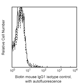
Profile of peripheral blood lymphocytes analyzed on a FACScan (BDIS, San Jose, CA)


BD Pharmingen™ Biotin Mouse Anti-Human PCNA

Regulatory Statusの凡例
Any use of products other than the permitted use without the express written authorization of Becton, Dickinson and Company is strictly prohibited.
Preparation and Storage
推奨アッセイ手順
1. Harvest, count and pellet cells following standard procedures.
Note: PCNA is expressed by proliferating cells. Using resting cells (eg, unstimulated PBMC) may give negative results.
2. While vortexing, add 5 ml cold 70% - 80% ethanol dropwise into the cell pellet (1-5 x 10e7 cells). Incubate at -20°C for at least 2 hours. These fixed cells can be stored at -20°C for up to 60 days prior to staining.
3. Wash twice with 30-40 ml staining buffer (PBS with 1% FBS, 0.09% NaN3), centrifuge for 10 minutes at 200g.
4. Resuspend the cells to a concentration of 1 X 10e7/ml.
5. Transfer 100 µl (1 X 10e6 cells) cell suspension into each sample tube.
6. Add 20 µl of properly diluted anti-PCNA antibody according to the protocol into the tubes above. Mix gently.
7. Incubate the tubes at room temperature (RT) for 20-30 minutes in the dark.
8. Wash with 2 ml of staining buffer at 200g for 5 minutes.
9. Aspirate the supernatant.
10. If using directly conjugated anti-PCNA, proceed to step 13.
11. If using purified anti-PCNA, add 50 µl of diluted secondary antibody (eg, cat. no. 555988), if using Biotin conjugated anti-PCNA, add 50µl of SAV-PE (Cat. No. 554061), to each sample tube and incubate at RT for 30 minutes in the dark.
12. Repeat steps 8 & 9.
13. Add 0.5 ml of staining buffer to each tube. If using FITC conjugated anti-PCNA or secondary antibody, add 10 µl of Propidium Iodide Staining Solution (Cat. No. 556463) to each tube; for PE conjugated anti-PCNA or secondary antibody, add 20 µl BD Via-Probe™ Cell Viability Solution (Cat. No. 555816) to each tube.
14. Proceed to flow cytometric analysis.
Product Notices
- Since applications vary, each investigator should titrate the reagent to obtain optimal results.
- Please refer to www.bdbiosciences.com/us/s/resources for technical protocols.
- Caution: Sodium azide yields highly toxic hydrazoic acid under acidic conditions. Dilute azide compounds in running water before discarding to avoid accumulation of potentially explosive deposits in plumbing.
関連製品

.png?imwidth=320)
The Proliferating Cell Nuclear Antigen (PCNA) was initially identified as a nuclear antigen in proliferating cells and was subsequently described as a subunit for DNA polymerase δ. PCNA protein levels peak during the S-phase of the cell cycle, at which time it forms a complex with the p21 inhibitor. PCNA is almost undetectable in other phases of the cycle. Because of its unique expression, PCNA has been extensively used in studies associating the prognosis of tumor progression and neoplastic proliferation. Human PCNA has been reported to be 262 amino acids with an apparent molecular weight of 36 kDa.
This antibody is routinely tested by flow cytometric analysis. Other applications were tested at BD Biosciences Pharmingen during antibody development only or reported in the literature.
Development References (6)
-
Garcia RL, Coltrera MD, Gown AM. Analysis of proliferative grade using anti-PCNA/cyclin monoclonal antibodies in fixed, embedded tissues. Comparison with flow cytometric analysis. Am J Pathol. 1989; 134(4):733-739. (Clone-specific: Flow cytometry). View Reference
-
Guesdon JL, Ternynck T, Avrameas S. The use of avidin-biotin interaction in immunoenzymatic techniques. J Histochem Cytochem. 1979; 27(8):1131-1139. (Biology). View Reference
-
Landberg G, Tan EM, Roos G. Flow cytometric multiparameter analysis of proliferating cell nuclear antigen/cyclin and Ki-67 antigen: a new view of the cell cycle. Exp Cell Res. 1990; 187(1):111-118. (Clone-specific: Flow cytometry). View Reference
-
Mathews MB, Bernstein RM, Franza BR Jr, Garrels JI. Identity of the proliferating cell nuclear antigen and cyclin. Nature. 1984; 309(5966):374-376. (Clone-specific: Flow cytometry). View Reference
-
Ogata K, Ogata Y, Nakamura RM, Tan EM. Purification and N-terminal amino acid sequence of proliferating cell nuclear antigen (PCNA)/cyclin and development of ELISA for anti-PCNA antibodies. J Immunol. 1985; 135(4):2623-2627. (Clone-specific: Flow cytometry). View Reference
-
Schlatt S, Weinbauer GF. Immunohistochemical localization of proliferating cell nuclear antigen as a tool to study cell proliferation in rodent and primate testes. Int J Androl. 1994; 17(4):214-222. (Clone-specific: Flow cytometry). View Reference
Please refer to Support Documents for Quality Certificates
Global - Refer to manufacturer's instructions for use and related User Manuals and Technical data sheets before using this products as described
Comparisons, where applicable, are made against older BD Technology, manual methods or are general performance claims. Comparisons are not made against non-BD technologies, unless otherwise noted.
For Research Use Only. Not for use in diagnostic or therapeutic procedures.
Report a Site Issue
This form is intended to help us improve our website experience. For other support, please visit our Contact Us page.