-
抗体試薬
- フローサイトメトリー用試薬
-
ウェスタンブロッティング抗体試薬
- イムノアッセイ試薬
-
シングルセル試薬
- BD® AbSeq Assay | シングルセル試薬
- BD Rhapsody™ Accessory Kits | シングルセル試薬
- BD® Single-Cell Multiplexing Kit | シングルセル試薬
- BD Rhapsody™ Targeted mRNA Kits | シングルセル試薬
- BD Rhapsody™ Whole Transcriptome Analysis (WTA) Amplification Kit | シングルセル試薬
- BD Rhapsody™ TCR/BCR Profiling Assays (VDJ Assays) | シングルセル試薬
- BD® OMICS-Guard Sample Preservation Buffer
-
細胞機能評価のための試薬
-
顕微鏡・イメージング用試薬
-
細胞調製・分離試薬
-
- BD® AbSeq Assay | シングルセル試薬
- BD Rhapsody™ Accessory Kits | シングルセル試薬
- BD® Single-Cell Multiplexing Kit | シングルセル試薬
- BD Rhapsody™ Targeted mRNA Kits | シングルセル試薬
- BD Rhapsody™ Whole Transcriptome Analysis (WTA) Amplification Kit | シングルセル試薬
- BD Rhapsody™ TCR/BCR Profiling Assays (VDJ Assays) | シングルセル試薬
- BD® OMICS-Guard Sample Preservation Buffer
- Japan (Japanese)
-
Change country/language
Old Browser
Looks like you're visiting us from {countryName}.
Would you like to stay on the current country site or be switched to your country?


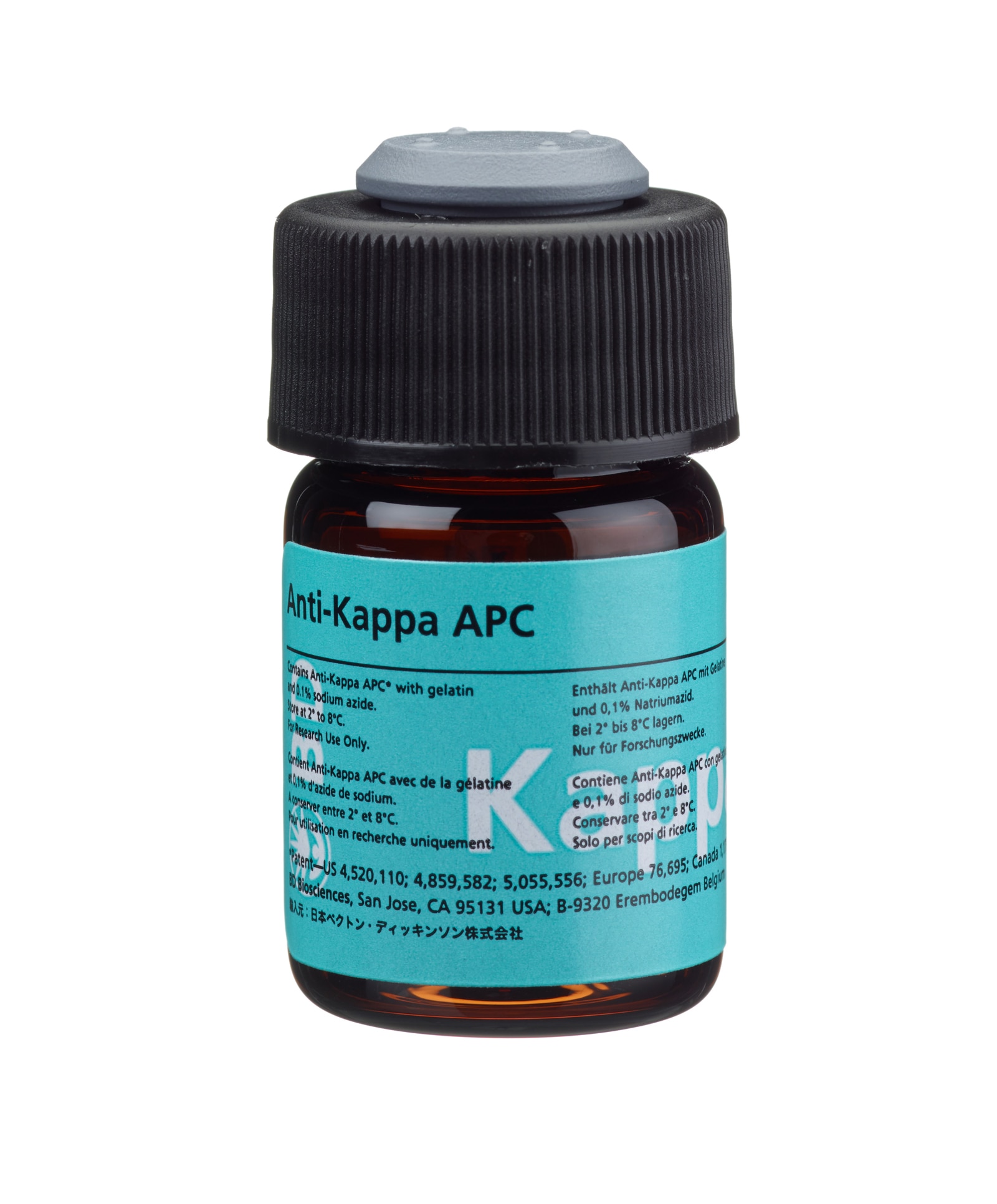
APC Mouse Anti-Human Kappa
Regulatory Statusの凡例
Any use of products other than the permitted use without the express written authorization of Becton, Dickinson and Company is strictly prohibited.
Preparation and Storage
Store vials at 2°C–8°C. Conjugated forms should not be frozen. Protect from exposure to light. Each reagent is stable until the expiration date shown on the bottle label when stored as directed.
The Anti-Kappa antibody, clone TB28-2, is derived from the hybridization of P3-X63-AG8.653 mouse myeloma cells with spleen cells isolated from CB6F1/J (C57BL/6J × BALB/cJ) mice immunized with human IgG, κ myeloma protein. The Anti-Kappa antibody specifically recognizes kappa (κ) light chains of human immunoglobulins.

Development References (15)
-
Ault KA. Flow cytometric evaluation of normal and neoplastic B cells. In: Rose NR, Friedman H, Fahey JL. Rose NR, Friedman H, Fahey JL, ed. Manual of Clinical Laboratory Immunology. 3rd ed.. Washington, DC: American Society for Microbiology; 1986:247-253. View Reference
-
Centers for Disease Control. Update: universal precautions for prevention of transmission of human immunodeficiency virus, hepatitis B virus, and other bloodborne pathogens in healthcare settings. MMWR. 1988; 37:377-388. (Biology).
-
Clinical and Laboratory Standards Institute. 2005. (Biology).
-
Foon KA, Todd RF. Immunologic classification of leukemia and lymphoma.. Blood. 1986; 68(1):1-31. (Biology). View Reference
-
Harris NL, Data RE. The distribution of neoplastic and normal B-lymphoid cells in nodular lymphomas: use of an immunoperoxidase technique on frozen sections.. Hum Pathol. 1982; 13(7):610-7. (Biology). View Reference
-
Kubagawa H, Gathings WE, Levitt D, Kearney JF, Cooper MD. Immunoglobulin isotype expression of normal pre-B cells as determined by immunofluorescence.. J Clin Immunol. 1982; 2(4):264-9. (Biology). View Reference
-
Meis JM, Osborne BM, Butler JJ. A comparative marker study of large cell lymphoma, Hodgkin's disease, and true histiocytic lymphoma in paraffin-embedded tissue.. Am J Clin Pathol. 1986; 86(5):591-9. (Biology). View Reference
-
Picker LJ, Weiss LM, Medeiros LJ, Wood GS, Warnke RA. Immunophenotypic criteria for the diagnosis of non-Hodgkin's lymphoma.. Am J Pathol. 1987; 128(1):181-201. (Biology). View Reference
-
Smith BR, Weinberg DS, Robert NJ, et al. Circulating monoclonal B lymphocytes in non-Hodgkin's lymphoma.. N Engl J Med. 1984; 311(23):1476-81. (Biology). View Reference
-
Stetler-Stevenson M, Braylan RC. Flow cytometric analysis of lymphomas and lymphoproliferative disorders.. Semin Hematol. 2001; 38(2):111-23. (Biology). View Reference
-
Stites DP, Casavant CH, McHugh TM, et al. Flow cytometric analysis of lymphocyte phenotypes in AIDS using monoclonal antibodies and simultaneous dual immunofluorescence.. Clin Immunol Immunopathol. 1986; 38(2):161-77. (Biology). View Reference
-
Tubbs RR, Sheibani K, Weiss RA, Sebek BA, Deodhar SD. Tissue immunomicroscopic evaluation of monoclonality of B-cell lymphomas: comparison with cell suspension studies.. Am J Clin Pathol. 1981; 76(1):24-8. (Biology). View Reference
-
Têtu B, Manning JT, Ordóñez NG. Comparison of monoclonal and polyclonal antibodies directed against immunoglobulin light and heavy chains in non-Hodgkin's lymphoma.. Am J Clin Pathol. 1986; 85(1):25-31. (Biology). View Reference
-
Weinberg DS, Pinkus GS, Ault KA. Cytofluorometric detection of B cell clonal excess: a new approach to the diagnosis of B cell lymphoma.. Blood. 1984; 63(5):1080-7. (Biology). View Reference
-
van Dongen JJ, Lhermitte L, Böttcher S, et al. EuroFlow antibody panels for standardized n-dimensional flow cytometric immunophenotyping of normal, reactive and malignant leukocytes. Leukemia. 2012; 26(9):1908-1975. (Biology). View Reference
Please refer to Support Documents for Quality Certificates
Global - Refer to manufacturer's instructions for use and related User Manuals and Technical data sheets before using this products as described
Comparisons, where applicable, are made against older BD Technology, manual methods or are general performance claims. Comparisons are not made against non-BD technologies, unless otherwise noted.
For Research Use Only. Not for use in diagnostic or therapeutic procedures.
Although not required, these products are manufactured in accordance with Good Manufacturing Practices.
Report a Site Issue
This form is intended to help us improve our website experience. For other support, please visit our Contact Us page.