
Preparation And Storage
Recommended Assay Procedures
Note: For flow cytometric analysis of human cells, Alexa Fluor® 488 Mouse Anti- Stat6 (pY641) mAb 18 (Cat. No. 612600) is recommended.
Product Notices
- This reagent has been pre-diluted for use at the recommended Volume per Test. We typically use 1 × 10^6 cells in a 100-µl experimental sample (a test).
- Please refer to www.bdbiosciences.com/us/s/resources for technical protocols.
- The Alexa Fluor®, Pacific Blue™, and Cascade Blue® dye antibody conjugates in this product are sold under license from Molecular Probes, Inc. for research use only, excluding use in combination with microarrays, or as analyte specific reagents. The Alexa Fluor® dyes (except for Alexa Fluor® 430), Pacific Blue™ dye, and Cascade Blue® dye are covered by pending and issued patents.
- Alexa Fluor® is a registered trademark of Molecular Probes, Inc., Eugene, OR.
- Alexa Fluor® 488 fluorochrome emission is collected at the same instrument settings as for fluorescein isothiocyanate (FITC).
- Caution: Sodium azide yields highly toxic hydrazoic acid under acidic conditions. Dilute azide compounds in running water before discarding to avoid accumulation of potentially explosive deposits in plumbing.
- Source of all serum proteins is from USDA inspected abattoirs located in the United States.
- For fluorochrome spectra and suitable instrument settings, please refer to our Multicolor Flow Cytometry web page at www.bdbiosciences.com/colors.
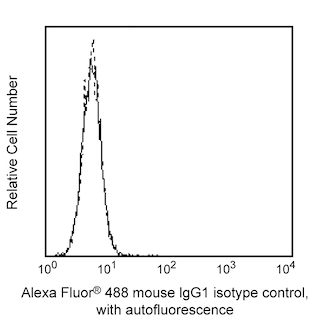
- An isotype control should be used at the same concentration as the antibody of interest.
Companion Products


STATs (signal transducers and activators of transcription) are critical mediators of the biologic activity of cytokines including Interleukins (IL) 2-5, IL-7, IL-15, GM-CSF, erythropoietin and growth hormone. Ligand-receptor interaction leads to activation of constitutively associated JAK family kinases and subsequent recruitment/activation of STATs by tyrosine phosphorylation. Active STATs then move to the nucleus to promote transcription of cytokine-inducible genes. Seven STAT proteins have been cloned, each of which is differentially expressed and/or activated in a cytokine-specific and cell type-specific manner. Stat6 plays an important role in signaling pathways that lead to the differentiation of T helper type 2 (Th2) cells from uncommitted CD4 T cell precursors. Moreover, IL-4, secreted by activated T lymphocytes, basophils, and mast cells, induces specific gene expression via the induction of tyrosine phosphorylation of Stat6 at tyrosine 641 (Y641). The SH3:SH2 domain of Stat6 associates with tyrosine-phosphorylated IL-4 receptor and the proximal Jak kinase phosphorylates Stat6 at Y641 on the C-terminal side of the SH2 domain. Stat6 is then released from the receptor, dimerizes, and is thought to contact the basal transcription machinery by binding to p300/CBP. While Stat6 is widely expressed in human tissues, it exhibits elevated expression in peripheral blood lymphocytes, colon, intestine, ovary, prostate, thymus, spleen, kidney, liver, lung, and placenta.
The J71-773.58.11 antibody recognizes mouse Stat6 phosphorylated at Y641.
Development References (8)
-
Bromberg J, Darnell JE. The role of STATs in transcriptional control and their impact on cellular function. Oncogene. 2000; 19(21):2468-2473. (Biology). View Reference
-
Daley JM, Brancato SK, Thomay AA, Reichner JS, Albina JE.. The phenotype of murine wound macrophages.. J Leukoc Biol. 2010; 87(1):59-67. (Clone-specific: Flow cytometry). View Reference
-
Dent AL, Hu-Li J, Paul WE, Staudt LM. T helper type 2 inflammatory disease in the absence of interleukin 4 and transcription factor STAT6. Proc Natl Acad Sci U S A. 1998; 95(23):13823-13828. (Biology). View Reference
-
Hale MB, Krutzik PO, Samra SS, Crane JM, Nolan GP.. Stage Dependent Aberrant Regulation of Cytokine-STAT Signaling in Murine Systemic Lupus Erythematosus. PLoS ONE. 2009; 4(8):e6756. (Clone-specific: Flow cytometry). View Reference
-
Heim MH. The Jak-STAT pathway: specific signal transduction from the cell membrane to the nucleus. Eur J Clin Invest. 1996; 26(1):1-12. (Biology). View Reference
-
Hou J, Schindler U, Henzel WJ, Ho TC, Brasseur M, McKnight SL. An interleukin-4-induced transcription factor: IL-4 Stat. Science. 1994; 265(5179):1701-1706. (Biology).
-
Mikita T, Campbell D, Wu P, Williamson K, Schindler U. Requirements for interleukin-4-induced gene expression and functional characterization of Stat6. Mol Cell Biol. 1996; 16(10):5811-5820. (Biology).
-
Quelle FW, Shimoda K, Thierfelder W, et al. Cloning of murine Stat6 and human Stat6, Stat proteins that are tyrosine phosphorylated in responses to IL-4 and IL-3 but are not required for mitogenesis. Mol Cell Biol. 1995; 15(6):3336-3343. (Biology).
Please refer to Support Documents for Quality Certificates
Global - Refer to manufacturer's instructions for use and related User Manuals and Technical data sheets before using this products as described
Comparisons, where applicable, are made against older BD Technology, manual methods or are general performance claims. Comparisons are not made against non-BD technologies, unless otherwise noted.
For Research Use Only. Not for use in diagnostic or therapeutic procedures.