
Preparation And Storage
Product Notices
- Since applications vary, each investigator should titrate the reagent to obtain optimal results.
- An isotype control should be used at the same concentration as the antibody of interest.
- Please refer to www.bdbiosciences.com/us/s/resources for technical protocols.
- The Alexa Fluor®, Pacific Blue™, and Cascade Blue® dye antibody conjugates in this product are sold under license from Molecular Probes, Inc. for research use only, excluding use in combination with microarrays, or as analyte specific reagents. The Alexa Fluor® dyes (except for Alexa Fluor® 430), Pacific Blue™ dye, and Cascade Blue® dye are covered by pending and issued patents.
- Alexa Fluor® 488 fluorochrome emission is collected at the same instrument settings as for fluorescein isothiocyanate (FITC).
- Alexa Fluor® is a registered trademark of Molecular Probes, Inc., Eugene, OR.
- For fluorochrome spectra and suitable instrument settings, please refer to our Multicolor Flow Cytometry web page at www.bdbiosciences.com/colors.
- Caution: Sodium azide yields highly toxic hydrazoic acid under acidic conditions. Dilute azide compounds in running water before discarding to avoid accumulation of potentially explosive deposits in plumbing.
Companion Products


.png?imwidth=320)
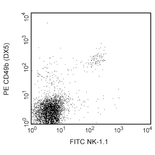
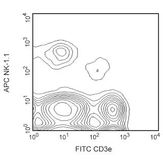
The ME-9F1 monoclonal antibody specifically binds to mouse CD146. The CD146 adhesion molecule is a type 1 transmembrane glycoprotein and member of the immunoglobulin superfamily. CD146 is expressed by blood vessel endothelial cells and may play roles in forming intercellular junctions between endothelial cells and influencing the transendothelial migration of other cell types. CD146 is also expressed by some melanoma cell lines, NK cells and neutrophils. CD146 is not detectable on mouse monocytes, dendritic cells, T cells, NKT cells, B cells and smooth muscle cells. Increased expression of CD146 is reportedly associated with NK cell maturation and may be used to characterize different functional NK cell subsets. Activated CD146-positive mouse NK cells reportedly are less cytotoxic and secrete less IFN-γ than their CD146-negative counterparts.
Development References (4)
-
Alais S, Allioli N, Pujades C, et al. HEMCAM/CD146 downregulates cell surface expression of beta1 integrins. J Cell Sci. 2001; 114(Pt 10):1847-1859. (Biology). View Reference
-
Despoix N, Walzer T, Jouve N, et al. Mouse CD146/MCAM is a marker of natural killer cell maturation. Eur J Immunol. 2008; 38(10):2855-2864. (Biology). View Reference
-
Schrage A, Loddenkemper C, Erben U, et al. Murine CD146 is widely expressed on endothelial cells and is recognized by the monoclonal antibody ME-9F1. Histochem Cell Biol. 2008; 129(4):441-451. (Clone-specific: Flow cytometry, Immunofluorescence, Immunohistochemistry, Immunoprecipitation). View Reference
-
Yang H, Wang S, Liu Z, et al. Isolation and characterization of mouse MUC18 cDNA gene, and correlation of MUC18 expression in mouse melanoma cell lines with metastatic ability. Gene. 2001; 265(1-2):133-145. (Biology). View Reference
Please refer to Support Documents for Quality Certificates
Global - Refer to manufacturer's instructions for use and related User Manuals and Technical data sheets before using this products as described
Comparisons, where applicable, are made against older BD Technology, manual methods or are general performance claims. Comparisons are not made against non-BD technologies, unless otherwise noted.
For Research Use Only. Not for use in diagnostic or therapeutic procedures.