

Preparation And Storage
Recommended Assay Procedures
Leukocytes are labeled with BD IMag™ anti-mouse CD8a Particles - DM according to the Magnetic Labeling Protocol. This labeled cell suspension is then placed within the magnetic field of BD IMag™ Cell Separation Magnet (Cat. No. 552311). Labeled cells migrate toward the magnet (positive fraction), leaving the unlabeled cells in suspension so they can be drawn off (negative fraction). The tube is then removed from the magnetic field for resuspension of the positive fraction. The separation is repeated twice to increase the purity of the positive fraction. The magnetic separation steps are diagrammed in the Separation Flow Chart. After the positive fraction is washed, the small size of the magnetic particles allows the positive fraction to be further evaluated in downstream applications such as flow cytometry.
MAGNETIC LABELING PROTOCOL
1. Prepare a single-cell suspension from the lymphoid tissue of interest according to standard laboratory procedures. Remove clumps of cells and/or debris by passing the suspension through a 70-µm nylon cell strainer.
2. Dilute BD IMag™ Buffer (10X) (Cat. No. 552362) 1:10 with sterile distilled water or prepare 1X BD IMag™ buffer by supplementing Phosphate Buffered Saline with 0.5% BSA, 2 mM EDTA, and 0.09% sodium azide. Place on ice.
Although our experience indicates that the use of Purified Rat Anti-Mouse CD16/CD32 (Mouse BD Fc Block™)(Cat. No. 553141/553142) is not required for optimal cell separation, some laboratories may want to use it in their studies. If adding Mouse BD Fc Block, proceed to Step 3. If not adding Mouse BD Fc Block, proceed to Step 4.
3. Add Mouse BD Fc Block at 0.25 µg/10^6 cells, and incubate on ice for 15 minutes.
4. Wash cells with at least an equal volume of 1X BD IMag buffer, and carefully aspirate all the supernatant.
5. Vortex the BD™ IMag anti-mouse CD8a Particles - DM thoroughly, and add 50 µl of particles for every 10^7 total cells.
6. MIX THOROUGHLY. Refrigerate at 6°C - 12°C for 30 minutes.
7. Bring the BD IMag-particle labeling volume up to 1 - 8 × 10^7 cells/ml with 1X BD IMag™ buffer, and immediately place the tube on the BD IMag™ Cell Separation Magnet. Incubate at room temperature for 6 - 8 minutes.
8. With the tube on the BD IMag™ Cell Separation Magnet, carefully aspirate off the supernatant. This supernatant contains the negative fraction.
9. Remove the tube from the BD IMag™ Cell Separation Magnet, and add 1X BD IMag buffer to the same volume as in Step 7. Gently resuspend cells by pipetting briefly, and return the tube to the BD IMag™ Cell Separation Magnet for another 2 - 4 minutes.
10. With the tube on the BD IMag™ Cell Separation Magnet, carefully aspirate off the supernatant and discard.
11. Repeat Steps 9 and 10.
12. After the final wash step, resuspend the positive fraction in an appropriate buffer and at an appropriate concentration for further analysis.
NOTE: Avoid nonspecific labeling by working quickly and adhering to recommended incubation times.
Product Notices
- Caution: Sodium azide yields highly toxic hydrazoic acid under acidic conditions. Dilute azide compounds in running water before discarding to avoid accumulation of potentially explosive deposits in plumbing.
- Source of all serum proteins is from USDA inspected abattoirs located in the United States.
- BD IMag™ particles are prepared from carboxy-functionalized magnetic particles which are manufactured by Skold Technology and are licensed under US patent number 7,169,618.
- Please refer to http://regdocs.bd.com to access safety data sheets (SDS).
- Please refer to www.bdbiosciences.com/us/s/resources for technical protocols.
Companion Products

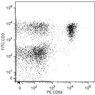
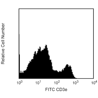
BD IMag™ anti-mouse CD8a Particles - DM are magnetic nanoparticles that have monoclonal antibody conjugated to their surfaces. These particles are optimized for the positive selection or depletion of CD8a-bearing leukocytes using the BD IMag™ Cell Separation Magnet. CD8a has been reported to be expressed on most thymocytes and a subpopulation of mature T lymphocytes (e.g. MHC class I-restricted T cells, including most T suppressor/cytotoxic cells). In addition, subsets of γδ TCR-bearing T cells, intestinal intraepithelial lymphocytes, and dendritic cells also have been reported to express CD8a.
Development References (10)
-
LeFrancois L. Extrathymic differentiation of intraepithelial lymphocytes: generation of a separate and unequal T-cell repertoire. Immunol Today. 1991; 12(12):436-438. (Biology). View Reference
-
Ledbetter JA, Herzenberg LA. Xenogeneic monoclonal antibodies to mouse lymphoid differentiation antigens. Immunol Rev. 1979; 47:63-90. (Biology). View Reference
-
Ledbetter JA, Rouse RV, Micklem HS, Herzenberg LA. T cell subsets defined by expression of Lyt-1,2,3 and Thy-1 antigens. Two-parameter immunofluorescence and cytotoxicity analysis with monoclonal antibodies modifies current views. J Exp Med. 1980; 152(2):280-295. (Biology). View Reference
-
MacDonald HR, Schreyer M, Howe RC, Bron C. Selective expression of CD8 alpha (Ly-2) subunit on activated thymic gamma/delta cells. Eur J Immunol. 1990; 20(4):927-930. (Biology). View Reference
-
Sydora BC, Brossay L, Hagenbaugh A, Kronenberg M, Cheroutre H. TAP-independent selection of CD8+ intestinal intraepithelial lymphocytes. J Immunol. 1996; 156(11):4209-4216. (Biology). View Reference
-
Traver D, Akashi K, Manz M, et al. Development of CD8alpha-positive dendritic cells from a common myeloid progenitor. Science. 2000; 290(5499):2152-2154. (Biology). View Reference
-
Vremec D, Zorbas M, Scollay R, et al. The surface phenotype of dendritic cells purified from mouse thymus and spleen: investigation of the CD8 expression by a subpopulation of dendritic cells. J Exp Med. 1992; 176(1):47-58. (Biology). View Reference
-
Walker ID, Murray BJ, Hogarth PM, Kelso A, McKenzie IF. Comparison of thymic and peripheral T cell Ly-2/3 antigens. Eur J Immunol. 1984; 14(10):906-910. (Biology). View Reference
-
Wang J, Klein JR. Thymus-neuroendocrine interactions in extrathymic T cell development. Science. 1994; 265(5180):1860-1862. (Biology). View Reference
-
van Ewijk W, van Soest PL, van den Engh GJ. Fluorescence analysis and anatomic distribution of mouse T lymphocyte subsets defined by monoclonal antibodies to the antigens Thy-1, Lyt-1, Lyt-2, and T-200. J Immunol. 1981; 127(6):2594-2604. (Biology). View Reference
Please refer to Support Documents for Quality Certificates
Global - Refer to manufacturer's instructions for use and related User Manuals and Technical data sheets before using this products as described
Comparisons, where applicable, are made against older BD Technology, manual methods or are general performance claims. Comparisons are not made against non-BD technologies, unless otherwise noted.
For Research Use Only. Not for use in diagnostic or therapeutic procedures.