BD Pharmingen™ PerCP Streptavidin
(RUO)

Preparation And Storage
Recommended Assay Procedures
PerCP has been reported to undergo significant photobleaching, the magnitude of which increases as laser power is increased or beam focus is narrowed. For tandem conjugates incorporating PerCP (e.g., PerCP-Cy5.5), the excitation and emission properties of PerCP and the kinetics of energy exchange between fluorochromes of the tandem dye may limit their effectiveness on high-speed and/or sorting flow cytometers. Therefore, for third-color flowcytometric analysis using ≥ 25-mW laser power, we recommend PE-Cy5 (formerly BD Cy-Chrome™)-conjugated reagents (eg, Cat. No. 554062).
Product Notices
- Since applications vary, each investigator should titrate the reagent to obtain optimal results.
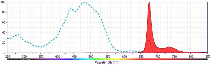
- For fluorochrome spectra and suitable instrument settings, please refer to our Multicolor Flow Cytometry web page at www.bdbiosciences.com/colors.
- PerCP is a photosynthetic accessory pigment from Glenodinium species of dinoflagellates, which is excited by the 488-nm light of an Argon ion laser and fluoresces at 675 nm. Therefore, PerCP-labelled antibodies can be used with FITC- and R-PE–labelled reagents in most single-laser flow cytometers with no significant spectral overlap of PerCP fluorescence with that of FITC or R-PE. PerCP has been reported to undergo significant photobleaching, the magnitude of which increases as laser power is increased or beam focus is narrowed. For third-color flow¬cytometric analysis using ≥25-mW laser power, we recommend PE-Cy5-, PE-Cy7–, or PerCP-Cy5.5-conjugated reagents.
- Caution: Sodium azide yields highly toxic hydrazoic acid under acidic conditions. Dilute azide compounds in running water before discarding to avoid accumulation of potentially explosive deposits in plumbing.
- Please refer to http://regdocs.bd.com to access safety data sheets (SDS).
- Cy is a trademark of GE Healthcare.
- Please refer to www.bdbiosciences.com/us/s/resources for technical protocols.
SAv-PerCP is a useful second-step reagent for the indirect immunofluorescent staining of cells in combination with biotinylated primary antibodies for flow cytometric analysis.
PerCP is a photosynthetic accessory pigment from Glenodinium species of dinoflagellates, which is excited by the 488-nm light of an Argon ion laser and fluoresces at 675 nm. Therefore, PerCP-labelled antibodies can be used with FITC- and R-PE-labelled reagents in most single-laser flow cytometers with no significant spectral overlap of PerCP fluorescence with that of FITC or R-PE.
Development References (4)
-
Afar B, Merrill J, Clark EA. Detection of lymphocyte subsets using three-color/single-laser flow cytometry and the fluorescent dye peridinin chlorophyll-alpha protein. J Clin Immunol. 1991; 11(5):254-261. (Biology). View Reference
-
Greimers R, Trebak M, Moutschen M, Jacobs N, Boniver J. Improved four-color flow cytometry method using fluo-3 and triple immunofluorescence for analysis of intracellular calcium ion ([Ca2+]i) fluxes among mouse lymph node B- and T-lymphocyte subsets. Cytometry. 1996; 23(3):205-217. (Biology). View Reference
-
Shapiro HM. Practical Flow Cytometry, 3rd Edition. New York: Wiley-Liss, Inc; 1995:280-281.
-
Waggoner AS, Ernst LA, Chen CH, Rechtenwald DJ. PE-CY5. A new fluorescent antibody label for three-color flow cytometry with a single laser. Ann N Y Acad Sci. 1993; 677:185-193. (Biology). View Reference
Please refer to Support Documents for Quality Certificates
Global - Refer to manufacturer's instructions for use and related User Manuals and Technical data sheets before using this products as described
Comparisons, where applicable, are made against older BD Technology, manual methods or are general performance claims. Comparisons are not made against non-BD technologies, unless otherwise noted.
For Research Use Only. Not for use in diagnostic or therapeutic procedures.