
Preparation And Storage
Recommended Assay Procedures
For optimal and reproducible results, BD Horizon Brilliant Stain Buffer should be used anytime two or more BD Horizon Brilliant dyes (including BD OptiBuild Brilliant reagents) are used in the same experiment. Fluorescent dye interactions may cause staining artifacts which may affect data interpretation. The BD Horizon Brilliant Stain Buffer was designed to minimize these interactions. More information can be found in the Technical Data Sheet of the BD Horizon Brilliant Stain Buffer (Cat. No. 563794).
Product Notices
- This antibody was developed for use in flow cytometry.
- The production process underwent stringent testing and validation to assure that it generates a high-quality conjugate with consistent performance and specific binding activity. However, verification testing has not been performed on all conjugate lots.
- Researchers should determine the optimal concentration of this reagent for their individual applications.
- An isotype control should be used at the same concentration as the antibody of interest.
- Caution: Sodium azide yields highly toxic hydrazoic acid under acidic conditions. Dilute azide compounds in running water before discarding to avoid accumulation of potentially explosive deposits in plumbing.
- For fluorochrome spectra and suitable instrument settings, please refer to our Multicolor Flow Cytometry web page at www.bdbiosciences.com/colors.
- Please refer to www.bdbiosciences.com/us/s/resources for technical protocols.
- BD Horizon Brilliant Stain Buffer is covered by one or more of the following US patents: 8,110,673; 8,158,444; 8,575,303; 8,354,239.
- BD Horizon Brilliant™ Violet 750 is covered by one or more of the following US patents: 8,158,444; 8,802,450; 8,575,303; 8,455,613; 8,227,187; 8,841,072; 8,110,673.
Companion Products


The OX-19 antibody reacts with CD5, a 69 kDa cell-surface glycoprotein found on thymocytes, peripheral T lymphocytes, and some thymic dendritic cells, but not on alloantigen-activated cytotoxic T cells, NK cells, γδ TCR-bearing intestinal intraepithelial lymphocytes, or dendritic epidermal T cells. A CD5+ subset of B lymphocytes has not been characterized in the rat. In the mouse and human, CD72 is the major ligand for CD5. While the OX-19 antibody is not mitogenic, its presence augments in vitro proliferative responses of T cells to lectins and allogeneic cells.
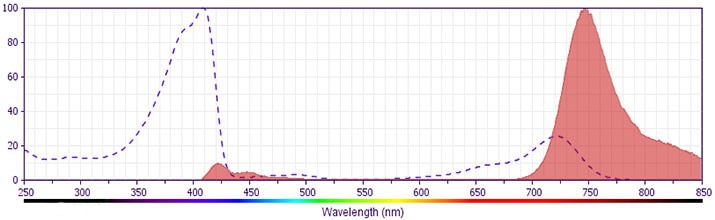
The antibody was conjugated to BD Horizon™ BV750 which is part of the BD Horizon Brilliant™ Violet family of dyes. This dye is a tandem fluorochrome of BD Horizon BV421 with an Ex Max of 405-nm and an acceptor dye with an Em Max at 750-nm. BD Horizon Brilliant BV750 can be excited by the violet laser (405 nm) and detected with a 750/30 nm filter with a 740 nm long pass. Due to spectral differences between labeled cells and beads, using BD™ CompBeads can result in incorrect spillover values when used with BD Horizon BV750 reagents. Therefore, the use of BD CompBeads or BD CompBeads Plus to determine spillover values for these reagents is not recommended.
Development References (8)
-
Bañuls MP, Alvarez A, Ferrero I, Zapata A, Ardavin C. Cell-surface marker analysis of rat thymic dendritic cells. Immunology. 1993; 79(2):298-304. (Clone-specific). View Reference
-
Luo W, Van de Velde H, von Hoegen I, Parnes JR, Thielemans K. Ly-1 (CD5), a membrane glycoprotein of mouse T lymphocytes and a subset of B cells, is a natural ligand of the B cell surface protein Lyb-2 (CD72). J Immunol. 1992; 148(6):1630-1634. (Biology). View Reference
-
Mallett S, Fossum S, Barclay AN. Characterization of the MRC OX40 antigen of activated CD4 positive T lymphocytes--a molecule related to nerve growth factor receptor. EMBO J. 1990; 9(4):1063-1068. (Clone-specific: (Co)-stimulation). View Reference
-
Paterson DJ, Jefferies WA, Green JR. Antigens of activated rat T lymphocytes including a molecule of 50,000 Mr detected only on CD4 positive T blasts. Mol Immunol. 1987; 24(12):1281-1290. (Immunogen). View Reference
-
Smith CA, Farrah T, Goodwin RG. The TNF receptor superfamily of cellular and viral proteins: activation, costimulation, and death. Cell. 1994; 76(6):959-962. (Biology). View Reference
-
Weinberg AD, Vella AT, Croft M. OX-40: life beyond the effector T cell stage. Semin Immunol. 1998; 10(6):471-480. (Clone-specific). View Reference
-
Weinberg AD, Wallin JJ, Jones RE, et al. Target organ-specific up-regulation of the MRC OX-40 marker and selective production of Th1 lymphokine mRNA by encephalitogenic T helper cells isolated from the spinal cord of rats with experimental autoimmune encephalomyelitis. J Immunol. 1994; 152(9):4712-4721. (Clone-specific). View Reference
-
al-Shamkhani A, Birkeland ML, Puklavec M, Brown MH, James W, Barclay AN. OX40 is differentially expressed on activated rat and mouse T cells and is the sole receptor for the OX40 ligand. Eur J Immunol. 1996; 26(8):1695-1699. (Clone-specific: Blocking). View Reference
Please refer to Support Documents for Quality Certificates
Global - Refer to manufacturer's instructions for use and related User Manuals and Technical data sheets before using this products as described
Comparisons, where applicable, are made against older BD Technology, manual methods or are general performance claims. Comparisons are not made against non-BD technologies, unless otherwise noted.
For Research Use Only. Not for use in diagnostic or therapeutic procedures.