Preparation And Storage
Recommended Assay Procedures
For optimal and reproducible results, BD Horizon Brilliant Stain Buffer should be used anytime two or more BD Horizon Brilliant dyes (including BD OptiBuild Brilliant reagents) are used in the same experiment. Fluorescent dye interactions may cause staining artifacts which may affect data interpretation. The BD Horizon Brilliant Stain Buffer was designed to minimize these interactions. More information can be found in the Technical Data Sheet of the BD Horizon Brilliant Stain Buffer (Cat. No. 563794).
Product Notices
- This antibody was developed for use in flow cytometry.
- The production process underwent stringent testing and validation to assure that it generates a high-quality conjugate with consistent performance and specific binding activity. However, verification testing has not been performed on all conjugate lots.
- Researchers should determine the optimal concentration of this reagent for their individual applications.
- An isotype control should be used at the same concentration as the antibody of interest.
- Caution: Sodium azide yields highly toxic hydrazoic acid under acidic conditions. Dilute azide compounds in running water before discarding to avoid accumulation of potentially explosive deposits in plumbing.
- For fluorochrome spectra and suitable instrument settings, please refer to our Multicolor Flow Cytometry web page at www.bdbiosciences.com/colors.
- Please refer to www.bdbiosciences.com/us/s/resources for technical protocols.
- BD Horizon Brilliant Stain Buffer is covered by one or more of the following US patents: 8,110,673; 8,158,444; 8,575,303; 8,354,239.
- BD Horizon Brilliant Ultraviolet 805 is covered by one or more of the following US patents: 8,110,673, 8,158,444; 8,227,187; 8,575,303; 8,354,239.
Companion Products


The OX-42 monoclonal antibody specifically binds to the CR3 complement (C3bi) receptor found on most monocytes, granulocytes, macrophages, dendritic cells, and microglia. It appears to recognize a common epitope shared by CD11b and CD11c (integrin αM and αX chains). OX-42 antibody inhibits C3bi binding activity.
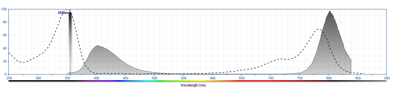
The antibody was conjugated to BD Horizon™ BUV805 which is part of the BD Horizon Brilliant™ Ultraviolet family of dyes. This dye is a tandem fluorochrome of BD Horizon BUV395 with an Ex Max of 348 nm and an acceptor dye with an Em Max at 805 nm. BD Horizon Brilliant BUV805 can be excited by the ultraviolet laser (355 nm) and detected with a 820/60 filter and a 770LP.
Development References (7)
-
Bañuls MP, Alvarez A, Ferrero I, Zapata A, Ardavin C. Cell-surface marker analysis of rat thymic dendritic cells. Immunology. 1993; 79(2):298-304. (Biology). View Reference
-
Dick AD, Ford AL, Forrester JV, Sedgwick JD. Flow cytometric identification of a minority population of MHC class II positive cells in the normal rat retina distinct from CD45lowCD11b/c+CD4low parenchymal microglia. J Leukoc Biol. 1995; 79(9):834-840. (Biology). View Reference
-
Ford AL, Goodsall AL, Hickey WF, Sedgwick JD. Normal adult ramified microglia separated from other central nervous system macrophages by flow cytometric sorting. Phenotypic differences defined and direct ex vivo antigen presentation to myelin basic protein-reactive CD4+ T cells compared. J Immunol. 1995; 154(9):4309-4321. (Biology). View Reference
-
Robinson AP, White TM, Mason DW. MRC OX-43: a monoclonal antibody which reacts with all vascular endothelium in the rat except that of brain capillaries. Immunology. 1986; 57(2):231-237. (Immunogen). View Reference
-
Robinson AP, White TM, Mason DW. Macrophage heterogeneity in the rat as delineated by two monoclonal antibodies MRC OX-41 and MRC OX-42, the latter recognizing complement receptor type 3. Immunology. 1986; 57(2):239-247. (Immunogen). View Reference
-
Shinoda M, Hoffer BJ, Olson L. Interactions of neurotrophic factors GDNF and NT-3, but not BDNF, with the immune system following fetal spinal cord transplantation. Brain Res. 1996; 722(1-2):153-167. (Biology). View Reference
-
Tamatani T, Kotani M, Miyasaka M. Characterization of the rat leukocyte integrin, CD11/CD18, by the use of LFA-1 subunit-specific monoclonal antibodies. Eur J Immunol. 1991; 21(3):627-633. (Biology). View Reference
Please refer to Support Documents for Quality Certificates
Global - Refer to manufacturer's instructions for use and related User Manuals and Technical data sheets before using this products as described
Comparisons, where applicable, are made against older BD Technology, manual methods or are general performance claims. Comparisons are not made against non-BD technologies, unless otherwise noted.
For Research Use Only. Not for use in diagnostic or therapeutic procedures.