Skip to main content Skip to navigation
PE Mouse Anti-Human LAG-3 (CD223)
Multicolor flow cytometric analysis of LAG-3 (CD223) expression on activated human peripheral blood lymphocytes. Human peripheral blood mononuclear cells were left untreated (Unstimulated Cells; Bottom Panel), or were cultured (Stimulated Cells; Top Panel) for 3 days with plate-bound Anti-Human CD3 (Cat. No. 555329; 10 µg/mL for coating) and soluble Anti-Human CD28 (Cat. No. 555725; 1 µg/mL) antibodies, and Human Recombinant IL-2 (Cat. No. 554603; 10 ng/mL). The Unstimulated and Stimulated Cells were pre-stained with BD Horizon Fixable Viability Stain 510 (FVS510; Cat. No. 564406) and fixed with BD Cytofix™ Fixation Buffer (Cat. No. 554655). The cells were then stained with Alexa Fluor® 647 Mouse Anti-Human CD8 (Cat. No. 557708), BD Horizon™ BV421 Mouse Anti-Human CD279 (PD-1) (Cat. No. 562516), and PE Mouse Anti-Human LAG-3 (CD223) (Cat. No. 565616/ 565617) antibodies. Two-color flow cytometric contour plots showing the correlated expression of LAG-3 (CD223) versus CD8 (Plots B and E) or CD279 (PD-1) (Plots C and F) were derived from live lymphocyte-gated events [ie, gated events with low level FVS510 incorporation and forward scattered-light signals (FSC-A) characteristic of intact lymphocytes; Plots A and D]. Flow cytometric analysis was performed using a BD LSRFortessa™ Flow Cytometer System.
Product Details
Down Arrow Up Arrow


BD Pharmingen™
LAG3; CD223; FDC; Lymphocyte activation gene 3 protein; Protein FDC
Human (QC Testing)
Mouse IgG1, κ
Human LAG-3 Recombinant Protein
Flow cytometry (Routinely Tested)
5 µl
3902
AB_2571727
Aqueous buffered solution containing BSA and ≤0.09% sodium azide.
RUO


Preparation And Storage

Store undiluted at 4°C and protected from prolonged exposure to light. Do not freeze. The monoclonal antibody was purified from tissue culture supernatant or ascites by affinity chromatography. The antibody was conjugated with R-PE under optimum conditions, and unconjugated antibody and free PE were removed.

Product Notices

  1. This reagent has been pre-diluted for use at the recommended Volume per Test. We typically use 1 × 10^6 cells in a 100-µl experimental sample (a test).
  2. Please refer to www.bdbiosciences.com/us/s/resources for technical protocols.
  3. Alexa Fluor® is a registered trademark of Molecular Probes, Inc., Eugene, OR.
  4. Caution: Sodium azide yields highly toxic hydrazoic acid under acidic conditions. Dilute azide compounds in running water before discarding to avoid accumulation of potentially explosive deposits in plumbing.
  5. For fluorochrome spectra and suitable instrument settings, please refer to our Multicolor Flow Cytometry web page at www.bdbiosciences.com/colors.
  6. Source of all serum proteins is from USDA inspected abattoirs located in the United States.
  7. An isotype control should be used at the same concentration as the antibody of interest.
565617 Rev. 2
Antibody Details
Down Arrow Up Arrow
T47-530

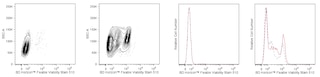
The T47-530 specifically recognizes the Lymphocyte Activation Gene 3 (LAG-3) protein which is also known as, Protein FDC, or CD223. LAG-3 is a ~70 kDa type I transmembrane glycoprotein that belongs to the Ig superfamily and exhibits homology to CD4. LAG-3 is expressed on NK cells, regulatory T cells, and activated conventional T cells with higher expression found on CD8+ T cells compared with CD4+ T cells. LAG-3 is an activation induced cell surface molecule that like CD4, binds MHC class II molecules, but with much higher affinity. This may enable LAG-3 to act as a negative competitor of CD4 for MHC class II ligand binding. LAG-3 may associate with the TCR-CD3 complex to downregulate TCR signal transduction and T cell clonal expansion. In contrast, LAG-3-induced signaling may promote dendritic cell activation.

565617 Rev. 2
Format Details
Down Arrow Up Arrow
PE
R-Phycoerythrin (PE), is part of the BD family of Phycobiliprotein dyes. This fluorochrome is a multimeric fluorescent phycobiliprotein with excitation maximum (Ex Max) of 496 nm and 566 nm and an emission maximum (Em Max) at 576 nm. PE is designed to be excited by the Blue (488 nm), Green (532 nm) and Yellow-Green (561 nm) lasers and detected using an optical filter centered near 575 nm (e.g., a 575/26-nm bandpass filter). As PE is excited by multiple lasers, this can result in cross-laser excitation and fluorescence spillover on instruments with various combinations of Blue, Green, and Yellow-Green lasers. Please ensure that your instrument’s configurations (lasers and optical filters) are appropriate for this dye.
altImg
PE
Yellow-Green 488 nm, 532 nm, 561 nm
496 nm, 566 nm
576 nm
565617 Rev.2
Citations & References
Down Arrow Up Arrow

Development References (5)

  1. Casati C, Camisaschi C, Novellino L, et al. Human lymphocyte activation gene-3 molecules expressed by activated T cells deliver costimulation signal for dendritic cell activation. J Immunol. 2008; 180(6):3782-3788. (Biology). View Reference
  2. Hannier S, Tournier M, Bismuth G, Triebel F. CD3/TCR complex-associated lymphocyte activation gene-3 molecules inhibit CD3/TCR signaling. J Immunol. 1998; 161(8):4058-4065. (Biology). View Reference
  3. Huang CT, Workman CJ, Flies D, et al. Role of LAG-3 in regulatory T cells. Immunity. 2004; 21(4):503-513. (Biology). View Reference
  4. Triebel F, Hacene K, Pichon MF. A soluble lymphocyte activation gene-3 (sLAG-3) protein as a prognostic factor in human breast cancer expressing estrogen or progesterone receptors. Cancer Lett. 2006; 235(1):147-153. (Biology). View Reference
  5. Triebel F, Jitsukawa S, Baixeras E, et al. LAG-3, a novel lymphocyte activation gene closely related to CD4. J Exp Med. 1990; 171(5):1393-1405. (Biology). View Reference
View All (5) View Less
565617 Rev. 2

Please refer to Support Documents for Quality Certificates


Global - Refer to manufacturer's instructions for use and related User Manuals and Technical data sheets before using this products as described


Comparisons, where applicable, are made against older BD Technology, manual methods or are general performance claims.  Comparisons are not made against non-BD technologies, unless otherwise noted.

For Research Use Only. Not for use in diagnostic or therapeutic procedures.