PE Mouse Anti-Human CD8
Clone RPA-T8 (RUO)
- Brand BD Pharmingen™
- Alternative Name CD8α; CD8A; CD8 alpha; Leu2; MAL; T8; p32
- Vol. Per Test 20 µl
- Isotype Mouse IgG1, κ
- Reactivity Human (QC Testing) Rhesus, Cynomolgus, Baboon (Tested in Development)
- Application
Flow cytometry (Routinely Tested)
- Immunogen Human CD8a
- Workshop No. IV T171; V T-CD08.03; VI 6T-CD8.1, 6T-081
- Entrez Gene ID 925
- Storage Buffer Aqueous buffered solution containing BSA, protein stabilizer, and ≤0.09% sodium azide.
- Regulatory Status RUO
Regulatory Status Legend
Description
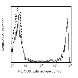
The RPA-T8 monoclonal antibody specifically binds to CD8 alpha (CD8á). CD8α is a type I transmembrane glycoprotein and a member of the immunoglobulin superfamily. CD8α is expressed by the majority of thymocytes, by subpopulations of αβ T cells and γδ T cells and by some NK cells. Cell surface CD8á is expressed either as a disulfide-linked homodimer (CD8αα) or as a heterodimer (CD8αâ) when disulfide-bonded to a CD8 beta chain (CD8â). CD8-positive αβ T cells coexpress both CD8áá homodimers and CD8αβ heterodimers whereas some γä T cells and NK cells express CD8αα homodimers. CD8 plays important roles in T cell activation and selection. The extracellular IgSF domain of CD8α binds to a non-polymorphic determinant on HLA class I molecules (α3 domain) and enables CD8 to function as a co-receptor with MHC class I-restricted TCR during T cell recognition of antigen. The cytoplasmic domain of CD8α associates with Lck, a Src family protein tyrosine kinase that is involved in intracellular signaling. The RPA-T8 and HIT8a monoclonal antibodies are not cross-blocking. This clone has been reported to react with a subset of peripheral blood lymphocytes, but not monocytes nor granuloyctes, of baboon and both rhesus and cynomolgus macaque monkey. In general, a higher frequency of CD8+ and CD4+CD8+ lymphocytes are observed in non-human primates compared to normal human donors.
Format
- Format PE
- Excitation Source Blue 488 nm,Green 532 nm,Yellow/Green 561 nm
- Excitation Max 496 nm
- Emission Max 578 nm
R-phycoerythrin (PE) is an accessory photosynthetic pigment found in red algae. It exists in vitro as a 240-kDa protein with 23 phycoerythrobilin chromophores per molecule. This makes PE one of the brightest fluorochromes for flow cytometry applications, but its photobleaching properties make it unsuitable for fluorescence microscopy.
Suggested Companion Products
PE Mouse IgG1, κ Isotype Control RUO
100 Tests
Cat No: 555749
Stain Buffer (FBS) RUO
500 mL
Cat No: 554656
Stain Buffer (BSA) RUO
500 mL
Cat No: 554657
Lysing Solution 10X Concentrate CE/IVD
100
Cat No: 349202
Lysing Buffer RUO
100 mL
Cat No: 555899
PE Mouse Anti-Human CD8 RPA-T8 RUO
25 Tests
Cat No: 561949
PE Mouse Anti-Human CD8 RPA-T8 RUO
50 Tests
Cat No: 557086
PE Mouse Anti-Human CD8 RPA-T8 RUO
500 Tests
Cat No: 561950
Resources & Tools | ||||||
---|---|---|---|---|---|---|
Spectrum Viewer | Panel Designer | Spectrum Viewer | Download TDS | Regulatory Document Website |
Preparation and Storage
Store undiluted at 4°C and protected from prolonged exposure to light. Do not freeze.
The monoclonal antibody was purified from tissue culture supernatant or ascites by affinity chromatography.
The antibody was conjugated with R-PE under optimum conditions, and unconjugated antibody and free PE were removed.
Product Notices
- This reagent has been pre-diluted for use at the recommended Volume per Test. We typically use 1 × 10^6 cells in a 100-µl experimental sample (a test).
- An isotype control should be used at the same concentration as the antibody of interest.
- Caution: Sodium azide yields highly toxic hydrazoic acid under acidic conditions. Dilute azide compounds in running water before discarding to avoid accumulation of potentially explosive deposits in plumbing.
- Source of all serum proteins is from USDA inspected abattoirs located in the United States.
- Species cross-reactivity detected in product development may not have been confirmed on every format and/or application.
- For fluorochrome spectra and suitable instrument settings, please refer to our Multicolor Flow Cytometry web page at www.bdbiosciences.com/colors.
- Please refer to www.bdbiosciences.com/us/s/resources for technical protocols.