-
Your selected country is
Middle East / Africa
- Change country/language
Old Browser
This page has been recently translated and is available in French now.
Looks like you're visiting us from {countryName}.
Would you like to stay on the current country site or be switched to your country?


Regulatory Status Legend
Any use of products other than the permitted use without the express written authorization of Becton, Dickinson and Company is strictly prohibited.
Preparation And Storage
Recommended Assay Procedures
BD™ CompBeads can be used as surrogates to assess fluorescence spillover (Compensation). When fluorochrome-conjugated antibodies are bound to BD CompBeads, they have spectral properties very similar to cells. However, for some fluorochromes there can be small differences in spectral emissions compared to cells, resulting in spillover values that differ when compared to biological controls. It is strongly recommended that when using a reagent for the first time, users compare the spillover on cells and BD CompBeads. This will ensure that BD CompBeads are appropriate for your specific cellular application.
For optimal and reproducible results, BD Horizon Brilliant Stain Buffer should be used anytime two or more BD Horizon Brilliant dyes are used in the same experiment. Fluorescent dye interactions may cause staining artifacts which may affect data interpretation. The BD Horizon Brilliant Stain Buffer was designed to minimize these interactions. More information can be found in the Technical Data Sheet of the BD Horizon Brilliant Stain Buffer (Cat. No. 563794/566349) or the BD Horizon Brilliant Stain Buffer Plus (Cat. No. 566385).
Note: When using high concentrations of antibody, background binding of this dye to erythroid cell subsets (mature erythrocytes and precursors) has been observed. For researchers studying these cell populations, or in cases where light scatter gating does not adequately exclude these cells from the analysis, this background may be an important factor to consider when selecting reagents for panel(s).
Product Notices
- The production process underwent stringent testing and validation to assure that it generates a high-quality conjugate with consistent performance and specific binding activity. However, verification testing has not been performed on all conjugate lots.
- An isotype control should be used at the same concentration as the antibody of interest.
- Caution: Sodium azide yields highly toxic hydrazoic acid under acidic conditions. Dilute azide compounds in running water before discarding to avoid accumulation of potentially explosive deposits in plumbing.
- For fluorochrome spectra and suitable instrument settings, please refer to our Multicolor Flow Cytometry web page at www.bdbiosciences.com/colors.
- BD Horizon Brilliant Stain Buffer is covered by one or more of the following US patents: 8,110,673; 8,158,444; 8,575,303; 8,354,239.
- CF™ is a trademark of Biotium, Inc.
- BD Horizon Brilliant Ultraviolet 563 is covered by one or more of the following US patents: 8,110,673; 8,158,444; 8,227,187; 8,575,303; 8,354,239.
- Please refer to http://regdocs.bd.com to access safety data sheets (SDS).
- Please refer to www.bdbiosciences.com/us/s/resources for technical protocols.
Companion Products


The WM53 monoclonal antibody specifically recognizes CD33 which is also known as Sialic acid-binding Ig-like lectin 3 (Siglec-3) or gp67. CD33 is a 67 kDa type I transmembrane glycoprotein that is variably expressed on myeloid progenitors, monocytes, macrophages, dendritic cells, neutrophils, basophils, mast cells, and on some activated T cells and NK cells. Normal lymphocytes, platelets, erythrocytes and pluripotent hematopoietic stem cells do not express the CD33 antigen. This glycoprotein reportedly functions as a sialic acid-dependent cell adhesion molecule and this function can be modulated by endogenous sialoglycoconjugates when CD33 is expressed on the membrane.
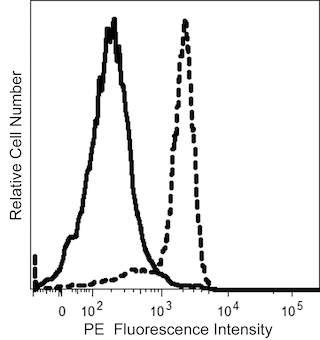
The antibody was conjugated to BD Horizon BUV563 which is part of the BD Horizon Brilliant™ Ultraviolet family of dyes. This dye is a tandem fluorochrome of BD Horizon BUV395 which has an Ex Max of 348 nm and an acceptor dye. The tandem has an Em Max at 563 nm. BD Horizon BUV563 can be excited by the 355 nm ultraviolet laser. On instruments with a 561 nm Yellow-Green laser, the recommended bandpass filter is 585/15 nm with a 535 nm long pass to minimize laser light leakage. When BD Horizon BUV563 is used with an instrument that does not have a 561 nm laser, a 560/40 nm filter with a 535 nm long pass may be more optimal. Due to the excitation and emission characteristics of the acceptor dye, there may be spillover into the PE and PE-CF594 detectors. However, the spillover can be corrected through compensation as with any other dye combination.

Development References (6)
-
Favaloro EJ, Bradstock KF, Kabral A, Grimsley P, Berndt MC. Characterization of monoclonal antibodies to the human myeloid-differentiation antigen, 'gp67' (CD-33). Dis Markers. 1987; 5(4):215-225. (Immunogen: Blocking, Flow cytometry, Fluorescence microscopy, Immunofluorescence, Immunoprecipitation, Radioimmunoassay). View Reference
-
Favaloro EJ, Bradstock KF, Kabral A, Grimsley P, Zowtyj H, Zola H. Further characterization of human myeloid antigens (gp160,95; gp150; gp67): investigation of epitopic heterogeneity and non-haemopoietic distribution using panels of monoclonal antibodies belonging to CD-11b, CD-13 and CD-33. Br J Haematol. 1988; 69(2):163-171. (Clone-specific: Blocking, Flow cytometry, Fluorescence microscopy, Immunofluorescence, Immunohistochemistry, Radioimmunoassay). View Reference
-
Favaloro EJ, Moraitis N, Koutts J, Exner T, Bradstock KF. Endothelial cells and normal circulating haemopoietic cells share a number of surface antigens. Thromb Haemost. 1989; 61(2):217-224. (Clone-specific: ELISA). View Reference
-
Freeman SD, Kelm S, Barber EK, Crocker PR. Characterization of CD33 as a new member of the sialoadhesin family of cellular interaction molecules. Blood. 1995; 85(8):2005-2012. (Biology). View Reference
-
Knapp W. W. Knapp .. et al., ed. Leucocyte typing IV : white cell differentiation antigens. Oxford New York: Oxford University Press; 1989:1-1182.
-
Nakamura Y, Noma M, Kidokoro M, et al. Expression of CD33 antigen on normal human activated T lymphocytes.. Blood. 1994; 83(5):1442-3. (Biology). View Reference
Please refer to Support Documents for Quality Certificates
Global - Refer to manufacturer's instructions for use and related User Manuals and Technical data sheets before using this products as described
Comparisons, where applicable, are made against older BD Technology, manual methods or are general performance claims. Comparisons are not made against non-BD technologies, unless otherwise noted.
For Research Use Only. Not for use in diagnostic or therapeutic procedures.
Report a Site Issue
This form is intended to help us improve our website experience. For other support, please visit our Contact Us page.