

Preparation And Storage
Product Notices
- Since applications vary, each investigator should titrate the reagent to obtain optimal results.
- Please refer to www.bdbiosciences.com/us/s/resources for technical protocols.
- The Alexa Fluor®, Pacific Blue™, and Cascade Blue® dye antibody conjugates in this product are sold under license from Molecular Probes, Inc. for research use only, excluding use in combination with microarrays, or as analyte specific reagents. The Alexa Fluor® dyes (except for Alexa Fluor® 430), Pacific Blue™ dye, and Cascade Blue® dye are covered by pending and issued patents.
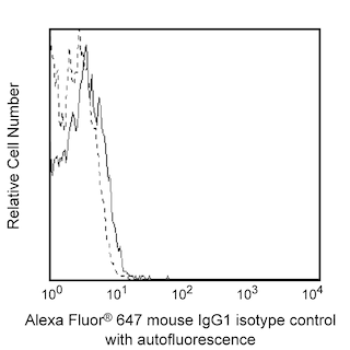
- Alexa Fluor® 647 fluorochrome emission is collected at the same instrument settings as for allophycocyanin (APC).
- Alexa Fluor® is a registered trademark of Molecular Probes, Inc., Eugene, OR.
- Caution: Sodium azide yields highly toxic hydrazoic acid under acidic conditions. Dilute azide compounds in running water before discarding to avoid accumulation of potentially explosive deposits in plumbing.
- For fluorochrome spectra and suitable instrument settings, please refer to our Multicolor Flow Cytometry web page at www.bdbiosciences.com/colors.
- An isotype control should be used at the same concentration as the antibody of interest.
Companion Products


The 54/HIF-1α monoclonal antibody specifically recognizes the alpha subunit (HIF-1alpha/HIF-1a/HIF1A) of the heterodimeric helix-loop-helix transcription factor, Hypoxia-induced factor 1 (HIF-1) complex which is involved in cellular O2 homeostasis. Under hypoxic conditions, HIF-1a accumulates and translocates into the nucleus, where it binds to the constitutively-expressed HIF-1 beta subunit (HIF-1b, also known as, Aryl hydrocarbon receptor nuclear translocator/ARNT). This complex can then bind to hypoxic response elements (HREs) of target genes, including glucose transporters, glycolytic enzymes, erythropoietin, vascular endothelial growth factor (VEGF), and various other genes that facilitate metabolic adaptation to hypoxia. HIF-1a is also known to be regulated by proinflammatory cytokines and bacterial products and plays an essential role in inflammation, immunity, and cancer. HIF-1a is rapidly degraded under normoxic conditions.
Development References (7)
-
Effects of cellular iron deficiency on the formation of vascular endothelial growth factor and angiogenesis. Iron deficiency and angiogenesis. Cancer Cell. 10(28)(Biology). View Reference
-
Fallone F, Britton S, Nieto L, Salles B, Muller C. ATR controls cellular adaptation to hypoxia through positive regulation of hypoxia-inducible factor 1 (HIF-1) expression. Oncogene. 2013; 32(37):4387-4396. (Biology). View Reference
-
Lee JW, Bae SH, Jeong JW, Kim SH, Kim KW. Hypoxia-inducible factor (HIF-1)alpha: its protein stability and biological functions.. Exp Mol Med. 2004; 36(1):1-12. (Biology). View Reference
-
Poitz DM, Augstein A, Weinert S, Braun-Dullaeus RC, Strasser RH, Schmeisser A. OxLDL and macrophage survival: essential and oxygen-independent involvement of the Hif-pathway.. Basic Res Cardiol. 2011; 106(5):761-72. (Biology). View Reference
-
Raval RR, Lau KW, Tran MG, et al. Contrasting properties of hypoxia-inducible factor 1 (HIF-1) and HIF-2 in von Hippel-Lindau-associated renal cell carcinoma.. Mol Cell Biol. 2005; 25(13):5675-86. (Clone-specific: Immunohistochemistry, Western blot). View Reference
-
Zagzag D, Lukyanov Y, Lan L, et al. Hypoxia-inducible factor 1 and VEGF upregulate CXCR4 in glioblastoma: implications for angiogenesis and glioma cell invasion.. Lab Invest. 2006; 86(12):1221-32. (Clone-specific: Immunofluorescence, Immunohistochemistry, Western blot). View Reference
-
van der Groep P, Bouter A, Menko FH, van der Wall E, van Diest PJ. High frequency of HIF-1alpha overexpression in BRCA1 related breast cancer.. Breast Cancer Res Treat. 2008; 111(3):475-80. (Clone-specific: Immunohistochemistry). View Reference
Please refer to Support Documents for Quality Certificates
Global - Refer to manufacturer's instructions for use and related User Manuals and Technical data sheets before using this products as described
Comparisons, where applicable, are made against older BD Technology, manual methods or are general performance claims. Comparisons are not made against non-BD technologies, unless otherwise noted.
For Research Use Only. Not for use in diagnostic or therapeutic procedures.