Preparation And Storage
Recommended Assay Procedures
For optimal and reproducible results, BD Horizon Brilliant Stain Buffer should be used anytime two or more BD Horizon Brilliant dyes (including BD OptiBuild Brilliant reagents) are used in the same experiment. Fluorescent dye interactions may cause staining artifacts which may affect data interpretation. The BD Horizon Brilliant Stain Buffer was designed to minimize these interactions. More information can be found in the Technical Data Sheet of the BD Horizon Brilliant Stain Buffer (Cat. No. 563794).
Product Notices
- This antibody was developed for use in flow cytometry.
- The production process underwent stringent testing and validation to assure that it generates a high-quality conjugate with consistent performance and specific binding activity. However, verification testing has not been performed on all conjugate lots.
- Researchers should determine the optimal concentration of this reagent for their individual applications.
- An isotype control should be used at the same concentration as the antibody of interest.
- Caution: Sodium azide yields highly toxic hydrazoic acid under acidic conditions. Dilute azide compounds in running water before discarding to avoid accumulation of potentially explosive deposits in plumbing.
- For fluorochrome spectra and suitable instrument settings, please refer to our Multicolor Flow Cytometry web page at www.bdbiosciences.com/colors.
- Please refer to www.bdbiosciences.com/us/s/resources for technical protocols.
- BD Horizon Brilliant Stain Buffer is covered by one or more of the following US patents: 8,110,673; 8,158,444; 8,575,303; 8,354,239.
- BD Horizon Brilliant Ultraviolet 805 is covered by one or more of the following US patents: 8,110,673, 8,158,444; 8,227,187; 8,575,303; 8,354,239.
Companion Products


Neurite adhesion molecule L1 has been implicated in neuron-neuron and neuron-Schwann cell adhesion in vertebrates. L1-like molecules, found in mouse, rat, chicken, and human, promote axonal elongation and may also play a role in regeneration of axons after injury. Molecular cloning data suggest 87% amino acid identity between mouse and human L1 molecules. 5G3 antigen (Ag), originally defined by monoclonal antibody 5G3, is considered to be the human homologue of mouse L1. The 5G3 antibody was developed against a human neuroblastoma cell line to use as a probe for the elucidating the biological characteristics of neuroblastoma. 5G3 specifically recognizes a neuroblastoma target glycoprotein antigen of 215 kDa and its 200 kDa precursor. The 215 kDa molecule is expressed on the cell surface; whereas the 200 kDa precursor is shed from the cell surface. The 215 and 200 kDa species also differ in their posttranslational modification patterns. The 5G3 antibody has been used as a marker for neuroblastoma, and to purify 5G3 Ag from normal adult human brain.
The antibody recognizes human L1 on human neuroblastoma cell lines and tissues. Reactivity has been tested on a variety of malignant and normal tissues. Squamous lung, squamous skin, and osteogenic sarcoma cell lines were positive, as were two out of eight melanoma cell lines tested. A variety of other cell lines and tumor tissues tested negative. 5G3 did not react with either T or B lymphoblastoid cell lines or a fibroblast cell line. Among all the normal tissues tested, mAb 5G3 reacted only with cerebellum.
The molecular masses observed using mAb 5G3 may vary among immunoprecipitation isolates. In normal human cerebellum, 5G3 Ag migrated as a 190/200 kDa doublet, 140 kDa band with minor bands at 80 and 65 kDa. 5G3 Ag isolated from SK-N-AS cells migrates as 200 to 215 kDa bands, or as a diffuse band ranging from 200 to 215 kDa. Additional bands have been described at 140 to 150 kDa in SK-N-AS cells. Only the 200 kDa band has been detected in culture media from SK-N-AS cells.
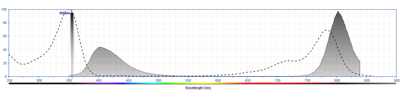
The antibody was conjugated to BD Horizon™ BUV805 which is part of the BD Horizon Brilliant™ Ultraviolet family of dyes. This dye is a tandem fluorochrome of BD Horizon BUV395 with an Ex Max of 348 nm and an acceptor dye with an Em Max at 805 nm. BD Horizon Brilliant BUV805 can be excited by the ultraviolet laser (355 nm) and detected with a 820/60 filter and a 770LP.
Development References (12)
-
Cheresh DA, Honsik CJ, Staffileno LK, Jung G, Reisfeld RA. Disialoganglioside GD3 on human melanoma serves as a relevant target antigen for monoclonal antibody-mediated tumor cytolysis. Proc Natl Acad Sci U S A. 1985; 82(15):5155-5159. (Biology). View Reference
-
Lemmon V, McLoon SC. The appearance of an L1-like molecule in the chick primary visual pathway. J Neurosci. 1986; 6(10):2987-2994. (Biology). View Reference
-
Mechtersheimer S, Gutwein P, Agmon-Levin N, et al. Ectodomain shedding of L1 adhesion molecule promotes cell migration by autocrine binding to integrins. J Cell Biol. 2001; 155(4):661-673. (Clone-specific: Western blot). View Reference
-
Mujoo K, Spiro RC, Reisfeld RA. Characterization of a unique glycoprotein antigen expressed on the surface of human neuroblastoma cells. J Biol Chem. 1986; 261(22):10299-10305. (Immunogen: Immunofluorescence, Immunohistochemistry, Western blot). View Reference
-
Nayeem N, Silletti S, Yang X, et al. A potential role for the plasmin(ogen) system in the posttranslational cleavage of the neural cell adhesion molecule L1. J Cell Sci. 1999; 112(24):4739-4749. (Clone-specific: Western blot). View Reference
-
Pancook JD, Reisfeld RA, Varki N, Vitiello A, Fox RI, Montgomery AM. Expression and regulation of the neural cell adhesion molecule L1 on human cells of myelomonocytic and lymphoid origin.. J Immunol. 1997; 158(9):4413-21. (Clone-specific). View Reference
-
Rathjen FG, Schachner M. Immunocytological and biochemical characterization of a new neuronal cell surface component (L1 antigen) which is involved in cell adhesion. EMBO J. 1984; 3(1):1-10. (Biology). View Reference
-
Rathjen FG, Wolff JM, Chang S, Bonhoeffer F, Raper JA. Neurofascin: a novel chick cell-surface glycoprotein involved in neurite-neurite interactions. Cell. 1987; 51(5):841-849. (Biology). View Reference
-
Reid RA, Hemperly JJ. Variants of human L1 cell adhesion molecule arise through alternate splicing of RNA. J Mol Neurosci. 1992; 3(3):127-135. (Biology). View Reference
-
Salton SR, Shelanski ML, Greene LA. Biochemical properties of the nerve growth factor-inducible large external (NILE) glycoprotein. J Neurosci. 1983; 3(12):2420-2430. (Biology). View Reference
-
Wolff JM, Frank R, Mujoo K, Spiro RC, Reisfeld RA, Rathjen FG. A human brain glycoprotein related to the mouse cell adhesion molecule L1.. J Biol Chem. 1988; 263(24):11943-7. (Clone-specific: Immunoaffinity chromatography). View Reference
-
Wolff R, Plow EF, Ginsberg MH. Interaction of thrombospondin with resting and stimulated human platelets. J Biol Chem. 1986; 261(15):6840-6846. (Clone-specific: Western blot). View Reference
Please refer to Support Documents for Quality Certificates
Global - Refer to manufacturer's instructions for use and related User Manuals and Technical data sheets before using this products as described
Comparisons, where applicable, are made against older BD Technology, manual methods or are general performance claims. Comparisons are not made against non-BD technologies, unless otherwise noted.
For Research Use Only. Not for use in diagnostic or therapeutic procedures.