
Preparation And Storage
Recommended Assay Procedures
For optimal and reproducible results, BD Horizon Brilliant Stain Buffer should be used anytime two or more BD Horizon Brilliant dyes (including BD OptiBuild Brilliant reagents) are used in the same experiment. Fluorescent dye interactions may cause staining artifacts which may affect data interpretation. The BD Horizon Brilliant Stain Buffer was designed to minimize these interactions. More information can be found in the Technical Data Sheet of the BD Horizon Brilliant Stain Buffer (Cat. No. 563794).
Product Notices
- This antibody was developed for use in flow cytometry.
- The production process underwent stringent testing and validation to assure that it generates a high-quality conjugate with consistent performance and specific binding activity. However, verification testing has not been performed on all conjugate lots.
- Researchers should determine the optimal concentration of this reagent for their individual applications.
- An isotype control should be used at the same concentration as the antibody of interest.
- Caution: Sodium azide yields highly toxic hydrazoic acid under acidic conditions. Dilute azide compounds in running water before discarding to avoid accumulation of potentially explosive deposits in plumbing.
- For fluorochrome spectra and suitable instrument settings, please refer to our Multicolor Flow Cytometry web page at www.bdbiosciences.com/colors.
- Please refer to www.bdbiosciences.com/us/s/resources for technical protocols.
- BD Horizon Brilliant Stain Buffer is covered by one or more of the following US patents: 8,110,673; 8,158,444; 8,575,303; 8,354,239.
- BD Horizon Brilliant Violet 421 is covered by one or more of the following US patents: 8,158,444; 8,362,193; 8,575,303; 8,354,239.
- Pacific Blue™ is a trademark of Molecular Probes, Inc., Eugene, OR.
Companion Products


The WT.5 monoclonal antibody specifically recognizes the alpha subunit of LFA-1 (αLβ2 integrin, CD11a/CD18), a heterodimeric surface glycoprotein which is found on the majority of leukocytes, but not on peritoneal macrophages or peritoneal mast cells. LFA-1 mediates a variety of heterotypic and homotypic intercellular adhesions through interaction with ICAM-1 (CD54) and ICAM-2 (CD102). WT.1 mAb recognizes both the activated and unactivated forms of LFA-1. It inhibits the binding of LFA-1 to ICAM-1 in several in vitro assays, including binding of Concanavalin A-stimulated lymphocytes (Con A blasts) to purified ICAM-1 and Mg2+-dependent aggregation of concanavalin A-stimulated blasts. It has also been reported to inhibit leukocyte infiltration in several in vivo models of inflammation.
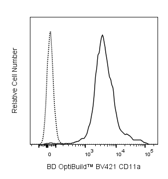
The antibody was conjugated to BD Horizon™ BV421 which is part of the BD Horizon Brilliant™ Violet family of dyes. With an Ex Max of 407-nm and Em Max at 421-nm, BD Horizon BV421 can be excited by the violet laser and detected in the standard Pacific Blue™ filter set (eg, 450/50-nm filter). BD Horizon BV421 conjugates are very bright, often exhibiting a 10 fold improvement in brightness compared to Pacific Blue conjugates.

Development References (10)
-
Bañuls MP, Alvarez A, Ferrero I, Zapata A, Ardavin C. Cell-surface marker analysis of rat thymic dendritic cells. Immunology. 1993; 79(2):298-304. (Clone-specific). View Reference
-
Fox CC, Jewell SD, Whitacre CC. Rat peritoneal mast cells present antigen to a PPD-specific T cell line. Cell Immunol. 1994; 158(1):253-264. (Clone-specific). View Reference
-
Kawasaki K, Yaoita E, Yamamoto T, Tamatani T, Miyasaka M, Kihara I. Antibodies against intercellular adhesion molecule-1 and lymphocyte function-associated antigen-1 prevent glomerular injury in rat experimental crescentic glomerulonephritis. J Immunol. 1993; 150(3):1074-1083. (Clone-specific). View Reference
-
Larson RS, Springer TA. Structure and function of leukocyte integrins. Immunol Rev. 1990; 114:181-217. (Biology). View Reference
-
Nishikawa K, Guo YJ, Miyasaka M, et al. Antibodies to intercellular adhesion molecule 1/lymphocyte function-associated antigen 1 prevent crescent formation in rat autoimmune glomerulonephritis. J Exp Med. 1993; 177(3):667-677. (Clone-specific). View Reference
-
Springer TA. Traffic signals for lymphocyte recirculation and leukocyte emigration: the multistep paradigm. Cell. 1994; 76(2):301-314. (Clone-specific). View Reference
-
Tamatani T, Kotani M, Miyasaka M. Characterization of the rat leukocyte integrin, CD11/CD18, by the use of LFA-1 subunit-specific monoclonal antibodies. Eur J Immunol. 1991; 21(3):627-633. (Immunogen). View Reference
-
Wada J, Shikata K, Makino H, et al. The critical role of intercellular adhesion molecule-1 in Masugi nephritis in rats. Nephron. 1996; 73(2):264-272. (Clone-specific). View Reference
-
Watanabe T, Arakawa T, Fukuda T, Higuchi K, Kobayashi K. Role of neutrophils in a rat model of gastric ulcer recurrence caused by interleukin-1 beta. Am J Pathol. 1997; 150(3):971-979. (Clone-specific). View Reference
-
Yamazaki T, Seko Y, Tamatani T, et al. Expression of intercellular adhesion molecule-1 in rat heart with ischemia/reperfusion and limitation of infarct size by treatment with antibodies against cell adhesion molecules. Am J Pathol. 1993; 143(2):410-418. (Clone-specific). View Reference
Please refer to Support Documents for Quality Certificates
Global - Refer to manufacturer's instructions for use and related User Manuals and Technical data sheets before using this products as described
Comparisons, where applicable, are made against older BD Technology, manual methods or are general performance claims. Comparisons are not made against non-BD technologies, unless otherwise noted.
For Research Use Only. Not for use in diagnostic or therapeutic procedures.