
Preparation And Storage
Recommended Assay Procedures
For optimal and reproducible results, BD Horizon Brilliant Stain Buffer should be used anytime two or more BD Horizon Brilliant dyes are used in the same experiment. Fluorescent dye interactions may cause staining artifacts which may affect data interpretation. The BD Horizon Brilliant Stain Buffer was designed to minimize these interactions. More information can be found in the Technical Data Sheet of the BD Horizon Brilliant Stain Buffer (Cat. No. 563794/566349) or the BD Horizon Brilliant Stain Buffer Plus (Cat. No. 566385).
Note: When using high concentrations of antibody, background binding of this dye to erythroid cell subsets (mature erythrocytes and precursors) has been observed. For researchers studying these cell populations, or in cases where light scatter gating does not adequately exclude these cells from the analysis, this background may be an important factor to consider when selecting reagents for panel(s).
Product Notices
- This reagent has been pre-diluted for use at the recommended Volume per Test. We typically use 1 × 10^6 cells in a 100-µl experimental sample (a test).
- An isotype control should be used at the same concentration as the antibody of interest.
- Caution: Sodium azide yields highly toxic hydrazoic acid under acidic conditions. Dilute azide compounds in running water before discarding to avoid accumulation of potentially explosive deposits in plumbing.
- For fluorochrome spectra and suitable instrument settings, please refer to our Multicolor Flow Cytometry web page at www.bdbiosciences.com/colors.
- BD Horizon Brilliant Ultraviolet 496 is covered by one or more of the following US patents: 8,110,673; 8,158,444; 8,227,187; 8,575,303; 8,354,239.
- BD Horizon Brilliant Stain Buffer is covered by one or more of the following US patents: 8,110,673; 8,158,444; 8,575,303; 8,354,239.
- Please refer to http://regdocs.bd.com to access safety data sheets (SDS).
- Species cross-reactivity detected in product development may not have been confirmed on every format and/or application.
- Please refer to www.bdbiosciences.com/us/s/resources for technical protocols.
Companion Products


The RPA-T8 monoclonal antibody specifically binds to CD8 alpha (CD8α). CD8α is a type I transmembrane glycoprotein and a member of the immunoglobulin superfamily. CD8α is expressed by the majority of thymocytes, by subpopulations of αβ T cells and γδ T cells and by some NK cells. Cell surface CD8α is expressed either as a disulfide-linked homodimer (CD8αα) or as a heterodimer (CD8αβ) when disulfide-bonded to a CD8 beta chain (CD8β). CD8-positive αβ T cells coexpress both CD8αα homodimers and CD8αβ heterodimers whereas some γδ T cells and NK cells express CD8αα homodimers. CD8 plays important roles in T cell activation and selection. The extracellular IgSF domain of CD8α binds to a non-polymorphic determinant on HLA class I molecules (α3 domain) and enables CD8 to function as a co-receptor with MHC class I-restricted TCR during T cell recognition of antigen. The cytoplasmic domain of CD8α associates with Lck, a Src family protein tyrosine kinase that is involved in intracellular signaling. The RPA-T8 and HIT8a monoclonal antibodies are not cross-blocking. This clone has been reported to react with a subset of peripheral blood lymphocytes, but not monocytes nor granuloyctes, of baboon and both rhesus and cynomolgus macaque monkey. In general, a higher frequency of CD8+ and CD4+CD8+ lymphocytes are observed in non-human primates compared to normal human donors.
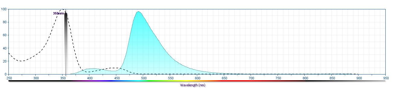
The antibody was conjugated to BD Horizon BUV496 which is part of the BD Horizon Brilliant™ Ultraviolet family of dyes. This dye is a tandem fluorochrome of BD Horizon BUV395 with an Ex Max of 348-nm and an acceptor dye with an Em Max at 496-nm. BD Horizon BUV496 can be excited by the ultraviolet laser (355 nm) and detected with a 515/30 nm filter with a 450LP. Due to the excitation of the acceptor dye by other laser lines, there may be significant spillover into the channel detecting BD Horizon V500 or BV510 (eg, 525/40-nm filter). However, the spillover can be corrected through compensation as with any other dye combination.
Development References (6)
-
Garbrecht F, Loebel A, Disanto JP, Flomenberg N. Chatacterization of Workshop antiCD8 mAb using human CD8-expressing murine L-cell transfectants. In: Schlossman SF. Stuart F. Schlossman .. et al., ed. Leucocyte typing V : white cell differentiation antigens : proceedings of the fifth international workshop and conference held in Boston, USA, 3-7 November, 1993. Oxford: Oxford University Press; 1995:354-356.
-
Jeong SH, Qiao M, Nascimbeni M, et al. Immunization with hepatitis C virus-like particles induces humoral and cellular immune responses in nonhuman primates. J Virol. 2004; 78(13):6995-7003. (Clone-specific: Flow cytometry). View Reference
-
Moebius U. Cluster report: CD8. In: Knapp W. W. Knapp .. et al., ed. Leucocyte typing IV : white cell differentiation antigens. Oxford New York: Oxford University Press; 1989:342-343.
-
Qiu X, Audet J, Wong G, et al. Successful treatment of ebola virus-infected cynomolgus macaques with monoclonal antibodies.. Sci Transl Med. 2012; 4(138):138ra81. (Clone-specific: Flow cytometry). View Reference
-
Sopper S, Stahl-Hennig C, Demuth M, Johnston IC, Dorries R, ter Meulen V. Lymphocyte subsets and expression of differentiation markers in blood and lymphoid organs of rhesus monkeys. Cytometry. 1997; 29(4):351-362. (Clone-specific: Flow cytometry). View Reference
-
Takami T, Saio M, Nakayama T, Tanaka Y-U, Liu Y. T-cell Blind Panel: Immunihistochemical analysis reporyt. In: Kishimoto T. Tadamitsu Kishimoto .. et al., ed. Leucocyte typing VI : white cell differentiation antigens : proceedings of the sixth international workshop and conference held in Kobe, Japan, 10-14 November 1996. New York: Garland Pub.; 1997:88-92.
Please refer to Support Documents for Quality Certificates
Global - Refer to manufacturer's instructions for use and related User Manuals and Technical data sheets before using this products as described
Comparisons, where applicable, are made against older BD Technology, manual methods or are general performance claims. Comparisons are not made against non-BD technologies, unless otherwise noted.
For Research Use Only. Not for use in diagnostic or therapeutic procedures.