
Preparation And Storage
Product Notices
- Since applications vary, each investigator should titrate the reagent to obtain optimal results.
- An isotype control should be used at the same concentration as the antibody of interest.
- Caution: Sodium azide yields highly toxic hydrazoic acid under acidic conditions. Dilute azide compounds in running water before discarding to avoid accumulation of potentially explosive deposits in plumbing.
- Source of all serum proteins is from USDA inspected abattoirs located in the United States.
- For fluorochrome spectra and suitable instrument settings, please refer to our Multicolor Flow Cytometry web page at www.bdbiosciences.com/colors.
- Please refer to www.bdbiosciences.com/us/s/resources for technical protocols.
Companion Products


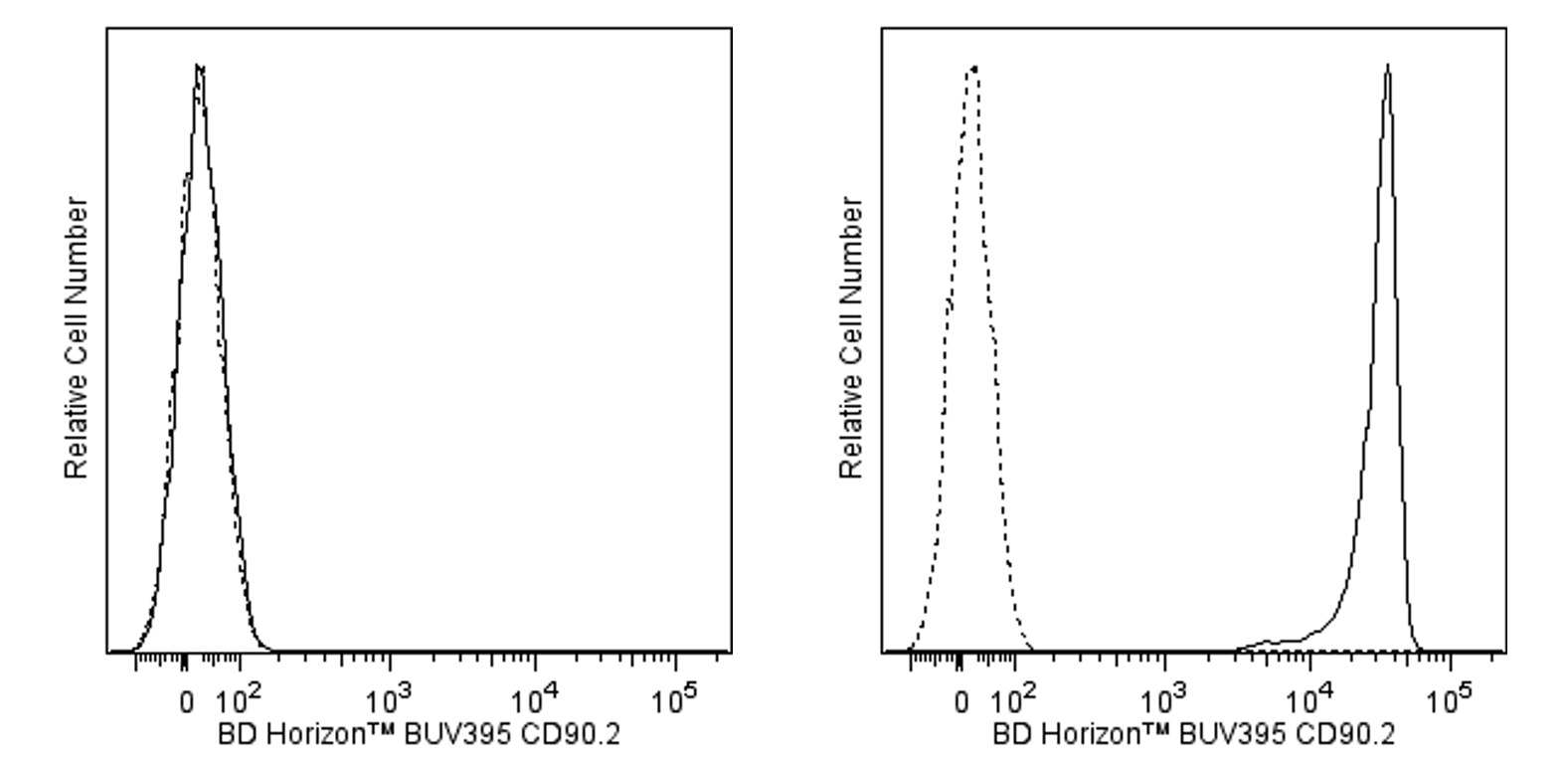
The 53-2.1 monoclonal antibody specifically binds to the CD90.2 (Thy-1.2) alloantigen on thymocytes, most peripheral T lymphocytes, some intraepithelial T lymphocytes (IEL, DEC), epithelial cells, fibroblasts, neurons, hematopoietic stem cells, but not B lymphocytes, of most mouse strains. The 53-2.1 antibody has been reported not to crossreact with Thy-1.1 (e.g., AKR/J, PL), or with rat Thy-1. CD90 is a glycophosphatidylinositol-anchored membrane glycoprotein of the Ig superfamily that is involved in signal transduction. In addition, there is evidence that CD90 mediates adhesion of thymocytes to thymic stroma. The 53-2.1 antibody has been reported to block the binding of the Rat Anti-Mouse CD90.2 antibody (Clone 30-H12) to immobilized thymocyte membranes.
The antibody was conjugated to BD Horizon BUV395 which has been exclusively developed by BD Biosciences as an optimal dye for use on a 355 nm laser equipped instrument. With an Ex Max at 348 nm and an Em Max at 395 nm, this dye has virtually no spillover into any other detector. BD Horizon BUV395 can be excited with a 355 nm laser and detected with a 379/28 filter.

Development References (12)
-
He HT, Naquet P, Caillol D, Pierres M. Thy-1 supports adhesion of mouse thymocytes to thymic epithelial cells through a Ca2(+)-independent mechanism. J Exp Med. 1991; 173(2):515-518. (Biology). View Reference
-
Hueber AO, Raposo G, Pierres M, He HT. Thy-1 triggers mouse thymocyte apoptosis through a bcl-2-resistant mechanism. J Exp Med. 1994; 179(3):785-796. (Biology). View Reference
-
Ikuta K, Uchida N, Friedman J, Weissman IL. Lymphocyte development from stem cells. Annu Rev Immunol. 1992; 10:759-783. (Biology). View Reference
-
Kroczek RA, Gunter KC, Germain RN, Shevach EM. Thy-1 functions as a signal transduction molecule in T lymphocytes and transfected B lymphocytes. Nature. 1986; 322(6075):181-184. (Biology). View Reference
-
LeFrancois L. Extrathymic differentiation of intraepithelial lymphocytes: generation of a separate and unequal T-cell repertoire. Immunol Today. 1991; 12(12):436-438. (Biology). View Reference
-
Ledbetter JA, Herzenberg LA. Xenogeneic monoclonal antibodies to mouse lymphoid differentiation antigens. Immunol Rev. 1979; 47:63-90. (Immunogen: Cytotoxicity, Flow cytometry, Radioimmunoassay). View Reference
-
Ledbetter JA, Rouse RV, Micklem HS, Herzenberg LA. T cell subsets defined by expression of Lyt-1,2,3 and Thy-1 antigens. Two-parameter immunofluorescence and cytotoxicity analysis with monoclonal antibodies modifies current views. J Exp Med. 1980; 152(2):280-295. (Clone-specific: Flow cytometry). View Reference
-
Radrizzani M, Carminatti H, Pivetta OH, Idoyaga Vargas VP. Developmental regulation of Thy 1.2 rate of synthesis in the mouse cerebellum. J Neurosci Res. 1995; 42(2):220-227. (Clone-specific). View Reference
-
Tigelaar RE, Lewis JM, Bergstresser PR. TCR gamma/delta+ dendritic epidermal T cells as constituents of skin-associated lymphoid tissue. J Invest Dermatol. 1990; 94(6):58S-63S. (Biology). View Reference
-
Williams AF, Gagnon J. Neuronal cell Thy-1 glycoprotein: homology with immunoglobulin. Science. 1982; 216(4547):696-703. (Biology). View Reference
-
Zheng B, Han S, Kelsoe G. T helper cells in murine germinal centers are antigen-specific emigrants that downregulate Thy-1. J Exp Med. 1996; 184(3):1083-1091. (Clone-specific: Immunohistochemistry). View Reference
-
Zhong RK, Donnenberg AD, Edison L, Harrison DE. The appearance of Thy-1- donor T cells in the peripheral circulation 3-6 weeks after bone marrow transplantation suggests an extrathymic origin. Int Immunol. 1996; 8(2):171-176. (Clone-specific: Flow cytometry). View Reference
Please refer to Support Documents for Quality Certificates
Global - Refer to manufacturer's instructions for use and related User Manuals and Technical data sheets before using this products as described
Comparisons, where applicable, are made against older BD Technology, manual methods or are general performance claims. Comparisons are not made against non-BD technologies, unless otherwise noted.
For Research Use Only. Not for use in diagnostic or therapeutic procedures.