Skip to main content Skip to navigation
APC-R700 Mouse Anti-Human CD13
Alert icon
Consider BD Horizon™ Red 718 Reagents, bright small molecule alternative to APC-R700 with lower spread into the APC detector. More Info #
APC-R700 Mouse Anti-Human CD13
Multiparameter flow cytometric analysis of CD13 expression on human peripheral blood leucocytes. Human whole blood was stained with either BD Horizon™ APC-R700 Mouse IgG1, κ Isotype Control (Cat. No. 564974; Left Panel) or BD Horizon APC-R700 Mouse Anti-Human CD13 antibody (Cat. No. 565124; Right Panel). Erythrocytes were lysed with BD FACS™ Lysing Solution (Cat. No. 349202). Two-parameter flow cytometric contour plots showing the correlated expression of CD13 (or Ig Isotype control staining) versus side light-scatter signals (SSC-A) were derived from gated events with the forward and side light-scatter characteristics of intact leucocyte populations. Flow cytometric analysis was performed using a BD™ LSR II Flow Cytometer System.
Product Details
Down Arrow Up Arrow


BD Horizon™
ANPEP; APN; Aminopeptidase N; Alanyl aminopeptidase; LAP1; PEPN
Human (QC Testing)
Mouse IgG1, κ
Human AML Cells
Flow cytometry (Routinely Tested)
5 µl
IV M44, M209
290
AB_2739076
Aqueous buffered solution containing BSA, protein stabilizer, glycerol and ≤0.09% sodium azide.
RUO


Preparation And Storage

Store undiluted at 4°C and protected from prolonged exposure to light. Do not freeze. The monoclonal antibody was purified from tissue culture supernatant or ascites by affinity chromatography. The antibody was conjugated to the dye under optimum conditions and unconjugated antibody and free dye were removed.

Recommended Assay Procedures

BD™ CompBeads can be used as surrogates to assess fluorescence spillover (Compensation).  When fluorochrome conjugated antibodies are bound to CompBeads, they have spectral properties very similar to cells.   However, for some fluorochromes there can be small differences in spectral emissions compared to cells, resulting in spillover values that differ when compared to biological controls.  It is strongly recommended that when using a reagent for the first time, users compare the spillover on cells and CompBead to ensure that BD Comp beads are appropriate for your specific cellular application.

Product Notices

  1. This reagent has been pre-diluted for use at the recommended Volume per Test. We typically use 1 × 10^6 cells in a 100-µl experimental sample (a test).
  2. An isotype control should be used at the same concentration as the antibody of interest.
  3. Source of all serum proteins is from USDA inspected abattoirs located in the United States.
  4. Alexa Fluor® is a registered trademark of Molecular Probes, Inc., Eugene, OR.
  5. Caution: Sodium azide yields highly toxic hydrazoic acid under acidic conditions. Dilute azide compounds in running water before discarding to avoid accumulation of potentially explosive deposits in plumbing.
  6. For fluorochrome spectra and suitable instrument settings, please refer to our Multicolor Flow Cytometry web page at www.bdbiosciences.com/colors.
  7. Please refer to http://regdocs.bd.com to access safety data sheets (SDS).
  8. Please refer to www.bdbiosciences.com/us/s/resources for technical protocols.
565124 Rev. 2
Antibody Details
Down Arrow Up Arrow
WM15

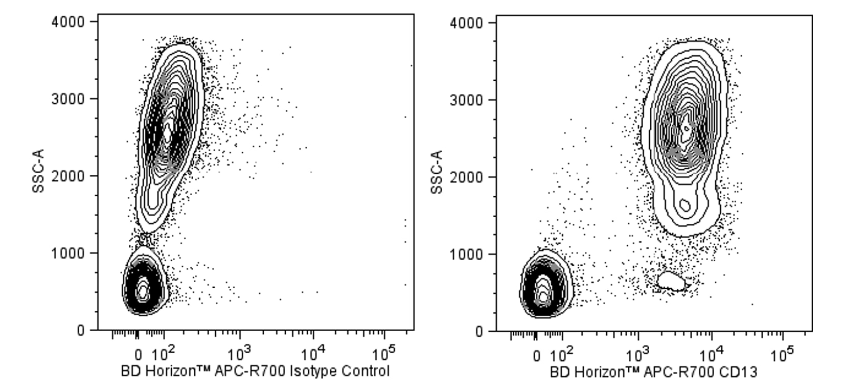
The WM15 monoclonal antibody specifically binds to CD13, the 150 kDa Type II integral membrane glycoprotein which is also known as aminopeptidase N. The CD13 antigen is the receptor for human coronavirus 229E, the causative agent for some cases of upper respiratory infection. This antibody binds to GM-progenitor cells, granulocytic and monocytic cells, and mast cells, but not to lymphocytes, platelets or erythrocytes. Aminopeptidase N is involved in the metabolism of many regulatory peptides.

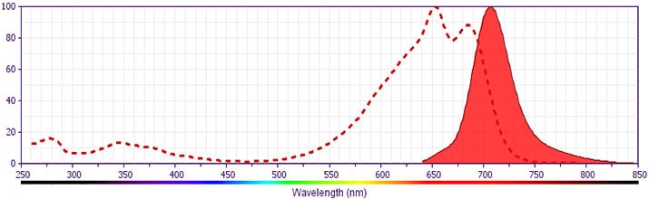
This antibody was conjugated to BD Horizon APC-R700, which has been developed exclusively by BD Biosciences as a better alternative to Alexa Fluor® 700. APC-R700 excites and emits at similar wavelengths to Alexa Fluor® 700 yet exhibits significantly improved brightness. This dye can be excited by the red laser and detected with the same filter set as Alexa Fluor® (eg, 730/45-nm filter).

565124 Rev. 2
Format Details
Down Arrow Up Arrow
APC-R700
The BD Horizon™ APC-R700 (APC-R700) Dye is a part of the BD APC red family of dyes. This tandem fluorochrome is comprised of an Allophycocyanin (APC) dye donor that has excitation maximum (Ex Max) of 651-nm and an acceptor dye, R700, with an emission maximum (Em Max) at 706-nm. APC-R700, driven by BD innovation, is designed to be excited by the red (627–640-nm) laser and detected using an optical filter centered near 710-nm (e.g., a 720/40-nm bandpass filter). APC-R700 is a brighter alternative to Alexa Fluor™ 700. Please ensure that your instrument’s configurations (lasers and optical filters) are appropriate for this dye.
altImg
APC-R700
Red 627-640 nm
651 nm
706 nm
565124 Rev.2
Citations & References
Down Arrow Up Arrow

Development References (8)

  1. Bradstock KF, Favaloro EJ, Kabral A, Kerr A, Hughes WG, Musgrove E. Myeloid progenitor surface antigen identified by monoclonal antibody.. Br J Haematol. 1985; 61(1):11-20. (Immunogen: Flow cytometry, Fluorescence activated cell sorting). View Reference
  2. Favaloro EJ, Bradstock KF, Kabral A, Grimsley P, Zowtyj H, Zola H. Further characterization of human myeloid antigens (gp160,95; gp150; gp67): investigation of epitopic heterogeneity and non-haemopoietic distribution using panels of monoclonal antibodies belonging to CD-11b, CD-13 and CD-33. Br J Haematol. 1988; 69(2):163-171. (Clone-specific: Blocking, Flow cytometry, Immunohistochemistry, Radioimmunoassay). View Reference
  3. Favaloro EJ, Moraitis N, Bradstock K, Koutts J. Co-expression of haemopoietic antigens on vascular endothelial cells: a detailed phenotypic analysis. Br J Haematol. 1990; 74(4):385-394. (Clone-specific: Flow cytometry). View Reference
  4. Favaloro EJ, Moraitis N, Koutts J, Exner T, Bradstock KF. Endothelial cells and normal circulating haemopoietic cells share a number of surface antigens. Thromb Haemost. 1989; 61(2):217-224. (Clone-specific: Flow cytometry, Immunohistochemistry). View Reference
  5. Gadd S. Cluster report: CD13. In: Knapp W. W. Knapp .. et al., ed. Leucocyte typing IV : white cell differentiation antigens. Oxford New York: Oxford University Press; 1989:782-784.
  6. Look AT, Ashmun RA, Shapiro LH, et al. Report on the CD13 (aminopeptidase N) cluster Workshop. In: Knapp W. W. Knapp .. et al., ed. Leucocyte typing IV : white cell differentiation antigens. Oxford New York: Oxford University Press; 1989:784-787.
  7. Yang P, Wang X. COVID-19: a new challenge for human beings. Cell Mol Immunol. 2020; 17(5):555-557. (Biology). View Reference
  8. Zola H. Leukocyte and stromal cell molecules : the CD markers. Hoboken, N.J.: Wiley-Liss; 2007.
View All (8) View Less
565124 Rev. 2

Please refer to Support Documents for Quality Certificates


Global - Refer to manufacturer's instructions for use and related User Manuals and Technical data sheets before using this products as described


Comparisons, where applicable, are made against older BD Technology, manual methods or are general performance claims.  Comparisons are not made against non-BD technologies, unless otherwise noted.

For Research Use Only. Not for use in diagnostic or therapeutic procedures.