
Preparation And Storage
Recommended Assay Procedures
For optimal and reproducible results, BD Horizon Brilliant Stain Buffer should be used anytime two or more BD Horizon Brilliant dyes (including BD Optibuild Brilliant reagents) are used in the same experiment. Fluorescent dye interactions may cause staining artifacts which may affect data interpretation. The BD Horizon Brilliant Stain Buffer was designed to minimize these interactions. More information can be found in the Technical Data Sheet of the BD Horizon Brilliant Stain Buffer (Cat. No. 563794/566349) or the BD Horizon Brilliant Stain Buffer Plus (Cat. No. 566385).
Product Notices
- This reagent has been pre-diluted for use at the recommended Volume per Test. We typically use 1 × 10^6 cells in a 100-µl experimental sample (a test).
- An isotype control should be used at the same concentration as the antibody of interest.
- Source of all serum proteins is from USDA inspected abattoirs located in the United States.
- Caution: Sodium azide yields highly toxic hydrazoic acid under acidic conditions. Dilute azide compounds in running water before discarding to avoid accumulation of potentially explosive deposits in plumbing.
- For fluorochrome spectra and suitable instrument settings, please refer to our Multicolor Flow Cytometry web page at www.bdbiosciences.com/colors.
- Cy is a trademark of GE Healthcare.
- BD Horizon Brilliant Stain Buffer is covered by one or more of the following US patents: 8,110,673; 8,158,444; 8,575,303; 8,354,239.
- BD Horizon Brilliant Violet 786 is covered by one or more of the following US patents: 8,110,673; 8,158,444; 8,227,187; 8,455,613; 8,575,303; 8,354,239.
- Please refer to www.bdbiosciences.com/us/s/resources for technical protocols.
Companion Products


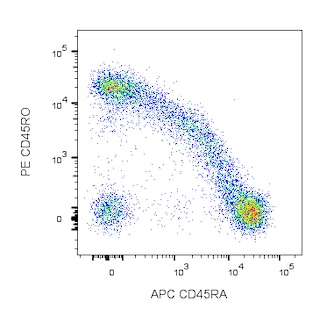
The UCHL1 monoclonal antibody specifically binds to the 180 kDa isoform of CD45 (aka, the Leukocyte Common Antigen). CD45RO is a type I transmembrane glycoprotein that has cytoplasmic protein tyrosine phosphatase activity and functions in signal transduction pathways. This CD45 isoform does not include amino acid sequences encoded by the variable CD45 exons A, B, or C. CD45RO is expressed on most thymocytes, activated T cells, memory T cells, granulocytes and monocytes, but only on a proportion of resting T cells. CD45RO and CD45RA antibodies seem to define complementary, predominantly non-overlapping, populations in resting peripheral T cells, demonstrating heterogeneity within the CD8 and CD4 subpopulations. CD45RO binds to CD22.
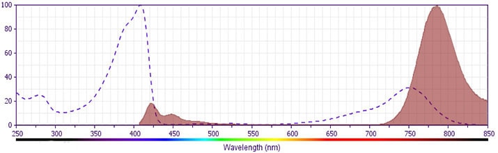
The antibody was conjugated to BD Horizon™ BV786 which is part of the BD Horizon Brilliant™ Violet family of dyes. This dye is a tandem fluorochrome of BD Horizon BV421 with an Ex Max of 405-nm and an acceptor dye with an Em Max at 786-nm. BD Horizon BV786 can be excited by the violet laser and detected in a filter used to detect Cy™7-like dyes (eg, 780/60-nm filter).
Development References (5)
-
Akbar AN, Terry L, Timms A, Beverley PC, Janossy G. Loss of CD45R and gain of UCHL1 reactivity is a feature of primed T cells. J Immunol. 1988; 140(7):2171-2178. (Clone-specific: Flow cytometry). View Reference
-
Knapp W. W. Knapp .. et al., ed. Leucocyte typing IV : white cell differentiation antigens. Oxford New York: Oxford University Press; 1989:1-1182.
-
Norton AJ, Ramsay AD, Smith SH, Beverley PC, Isaacson PG. Monoclonal antibody (UCHL1) that recognises normal and neoplastic T cells in routinely fixed tissues. J Clin Pathol. 1986; 39(4):399-405. (Immunogen: Immunohistochemistry). View Reference
-
Smith SH, Brown MH, Rowe D, Callard RE, Beverley PC. Functional subsets of human helper-inducer cells defined by a new monoclonal antibody, UCHL1. Immunology. 1986; 58(1):63-70. (Clone-specific: Flow cytometry, Immunohistochemistry, Immunoprecipitation). View Reference
-
Zola H. Leukocyte and stromal cell molecules : the CD markers. Hoboken, N.J.: Wiley-Liss; 2007.
Please refer to Support Documents for Quality Certificates
Global - Refer to manufacturer's instructions for use and related User Manuals and Technical data sheets before using this products as described
Comparisons, where applicable, are made against older BD Technology, manual methods or are general performance claims. Comparisons are not made against non-BD technologies, unless otherwise noted.
For Research Use Only. Not for use in diagnostic or therapeutic procedures.