
Preparation And Storage
Product Notices
- This reagent has been pre-diluted for use at the recommended Volume per Test. We typically use 1 × 10^6 cells in a 100-µl experimental sample (a test).
- Source of all serum proteins is from USDA inspected abattoirs located in the United States.
- An isotype control should be used at the same concentration as the antibody of interest.
- Please refer to www.bdbiosciences.com/us/s/resources for technical protocols.
- Caution: Sodium azide yields highly toxic hydrazoic acid under acidic conditions. Dilute azide compounds in running water before discarding to avoid accumulation of potentially explosive deposits in plumbing.
- For fluorochrome spectra and suitable instrument settings, please refer to our Multicolor Flow Cytometry web page at www.bdbiosciences.com/colors.
- Pacific Blue™ is a trademark of Molecular Probes, Inc., Eugene, OR.
- Brilliant Violet™ 421 is a trademark of Sirigen.
Companion Products


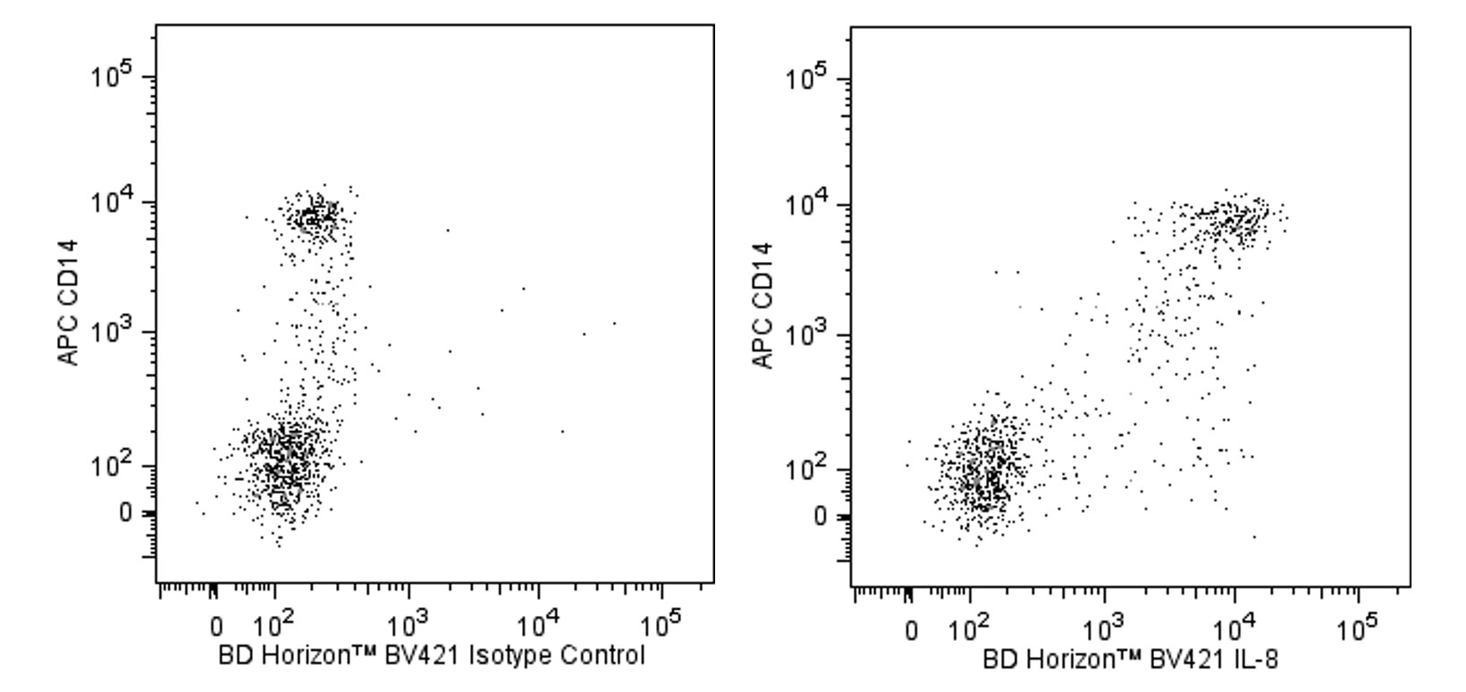
The G265-8 monoclonal antibody specifically binds to both the 72 and 77 amino acid isoforms of human Interleukin-8 (IL-8). IL-8 is secreted as an 8-9 kDa, non-glycosylated proinflammatory chemokine protein also known as chemokine (C-X-C motif) ligand 8 (CXCL8). IL-8 is synthesized as a 99 amino acid precursor that is proteolytically processed into several isoforms. The 72 amino acid isoform is produced by monocytes, macrophages, granulocytes, epithelial cells, and fibroblasts in response to pro-inflammatory stimuli including cytokines and microbial agents. It is also expressed by endothelial cells, fibroblasts, keratinocytes, lymphocytes, and a variety of tumor cells. In response to IL-4, IL-10 and TGFβ, the cellular production of IL-8 is inhibited. IL-8 is crucial for the activation and recruitment of neutrophils to inflammatory sites. IL-8 is also a chemoattractant for basophils and T-lymphocytes. IL-8 possesses angiogenic activity and can be associated with tumor angiogenesis and metastasis. The 77 amino acid IL-8 isoform is primarily produced by endothelial cells. This larger isoform is reportedly a less potent neutrophil activator than the 72 amino acid isoform. IL-8 binds to and signals through two G-protein-coupled receptors, IL-8RA (CXCR1/CD181) and IL-8RB (CXCR2/CD182).
The antibody was conjugated to BD Horizon™ BV421 which is part of the BD Horizon™ Brilliant Violet™ family of dyes. With an Ex Max of 407-nm and Em Max at 421-nm, BD Horizon™ BV421 can be excited by the violet laser and detected in the standard Pacific Blue™ filter set (eg, 450/50-nm filter). BD Horizon™ BV421 conjugates are very bright, often exhibiting a 10 fold improvement in brightness compared to Pacific Blue™ conjugates.

Development References (9)
-
Car BD, Meloni F, Luisetti M, Semenzato G, Gialdroni-Grassi G, Walz A. Elevated IL-8 and MCP-1 in the bronchoalveolar lavage fluid of patients with idiopathic pulmonary fibrosis and pulmonary sarcoidosis. Am J Respir Crit Care Med. 1994; 149(3 Pt 1):655-659. (Biology). View Reference
-
Emadi S, Clay D, Desterke C, et al. IL-8 and its CXCR1 and CXCR2 receptors participate in the control of megakaryocytic proliferation, differentiation, and ploidy in myeloid metaplasia with myelofibrosis. Blood. 2005; 105(2):464-473. (Clone-specific: Flow cytometry, Immunocytochemistry (cytospins), Immunofluorescence). View Reference
-
Hebert CA, Luscinskas FW, Kiely JM, et al. Endothelial and leukocyte forms of IL-8. Conversion by thrombin and interactions with neutrophils. J Immunol. 1990; 145(9):3033-3040. (Biology). View Reference
-
Larsen CG, Anderson AO, Appella E, Oppenheim JJ, Matsushima K. The neutrophil-activating protein (NAP-1) is also chemotactic for T lymphocytes. Science. 1989; 243(4897):1464-1466. (Biology). View Reference
-
Leonard EJ, Skeel A, Yoshimura T, Noer K, Kutvirt S, Van Epps D. Leukocyte specificity and binding of human neutrophil attractant/activation protein-1. J Immunol. 1990; 144(41323):1323-1330. (Biology). View Reference
-
Lizasa H, Matsushima K. IL-8. In: Oppenheim JJ, Feldmann M, Durum SK, Hirano T, Vilcek J, Nicola NA, ed. Cytokine Reference : A compendium of cytokines and other mediators of host defense. San Diego: Academic Press; 2001:1061-1067.
-
Matsushima K, Oppenheim JJ. Interleukin 8 and MCAF: novel inflammatory cytokines inducible by IL 1 and TNF. Cytokine. 1989; 1(1):2-13. (Biology). View Reference
-
Miller LS, Sorensen OE, Liu PT, et al. TGF-alpha regulates TLR expression and function on epidermal keratinocytes. J Immunol. 2005; 174(10):6137-6143. (Clone-specific: ELISA). View Reference
-
Prussin C, Metcalfe DD. Detection of intracytoplasmic cytokine using flow cytometry and directly conjugated anti-cytokine antibodies. J Immunol Methods. 1995; 188(1):117-128. (Biology). View Reference
Please refer to Support Documents for Quality Certificates
Global - Refer to manufacturer's instructions for use and related User Manuals and Technical data sheets before using this products as described
Comparisons, where applicable, are made against older BD Technology, manual methods or are general performance claims. Comparisons are not made against non-BD technologies, unless otherwise noted.
For Research Use Only. Not for use in diagnostic or therapeutic procedures.