
Preparation And Storage
Recommended Assay Procedures
CAUTION: B73.1 binding is inhibited by human serum or aggregated IgG. In whole blood preparations, CD16 shows variable reactivity with granulocytes.
Product Notices
- This reagent has been pre-diluted for use at the recommended Volume per Test. We typically use 1 × 10^6 cells in a 100-µl experimental sample (a test).
- An isotype control should be used at the same concentration as the antibody of interest.
- Source of all serum proteins is from USDA inspected abattoirs located in the United States.
- Please observe the following precautions: Absorption of visible light can significantly alter the energy transfer occurring in any tandem fluorochrome conjugate; therefore, we recommend that special precautions be taken (such as wrapping vials, tubes, or racks in aluminum foil) to prevent exposure of conjugated reagents, including cells stained with those reagents, to room illumination.
- Caution: Sodium azide yields highly toxic hydrazoic acid under acidic conditions. Dilute azide compounds in running water before discarding to avoid accumulation of potentially explosive deposits in plumbing.
- For fluorochrome spectra and suitable instrument settings, please refer to our Multicolor Flow Cytometry web page at www.bdbiosciences.com/colors.
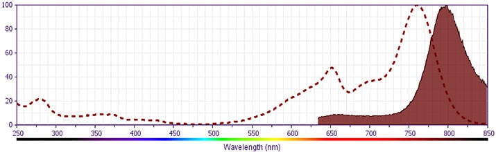
- BD APC-H7 is a tandem conjugate and an analog of APC-Cy7 with the same spectral properties. It has decreased intensity but it is engineered for greater stability and less spillover in the APC channel and consequently offers better performance than APC-Cy7. It has an absorption maximum of approximately 650 nm. When excited by light from a red laser, the APC fluorochrome can transfer energy to the cyanine dye, which then emits at a longer wavelength. The resulting fluorescent emission maximum is approximately 767 nm. BD recommends that a 750-nm longpass filter be used along with a red-sensitive detector such as the Hamamatsu R3896 PMT. As with APC-Cy7 special filters are required when using APC-H7 in conjunction with APC. Note: Although our APC-H7 products demonstrate higher lot-to lot consistency than other APC tandem conjugate products, and every effort is made to minimize the lot-to-lot variation in residual emission from APC, it is strongly recommended that every lot be tested for differences in the amount of compensation required and that individual compensation controls are run for each APC-H7 conjugate.
- Although BD APC-H7 is engineered to minimize spillover to the APC channel and is more stable and less affected by light, temperature, and formaldehyde-based fixatives, compared to other APC-cyanine tandem dyes, it is still good practice to minimize as much as possible, any light, temperature and fixative exposure when working with all fluorescent conjugates.
- Cy is a trademark of GE Healthcare.
- Please refer to www.bdbiosciences.com/us/s/resources for technical protocols.
Companion Products


The B73.1 monoclonal antibody specifically binds to CD16, the 50-70 kDa low affinity receptor for IgG, IgG Fc receptor III (FcγRIII). CD16 is expressed on NK cells, neutrophils, and on a subset of T cells from certain individuals. The B73.1 antibody binds to CD16-positive neutrophils with lower intensity when compared with some other CD16-specific antibodies. A variable number of CD16-positive lymphocytes coexpress either the CD57 antigen or low-density CD8 antigen or both. The B73.1 antibody can block Fc receptor functions mediated by CD16.
Development References (7)
-
Lanier LL, Kipps TJ, Phillips JH. Functional properties of a unique subset of cytotoxic CD3+ T lymphocytes that express Fc receptors for IgG (CD16/Leu-11 antigen). J Exp Med. 1985; 162(6):2089-2106. (Clone-specific: Flow cytometry). View Reference
-
Lanier LL, Le AM, Civin CI, Loken MR, Phillips JH. The relationship of CD16 (Leu-11) and Leu-19 (NKH-1) antigen expression on human peripheral blood NK cells and cytotoxic T lymphocytes. J Immunol. 1986; 136(12):4480-4486. (Biology). View Reference
-
Lanier LL, Le AM, Phillips JH, Warner NL, Babcock GF. Subpopulations of human natural killer cells defined by expression of the Leu-7 (HNK-1) and Leu-11 (NK-15) antigens. J Immunol. 1983; 131(4):1789-1796. (Biology). View Reference
-
Perussia B, Acuto O, Terhorst C, et al. Human natural killer cells analyzed by B73.1, a monoclonal antibody blocking Fc receptor functions. II. Studies of B73.1 antibody-antigen interaction on the lymphocyte membrane. J Immunol. 1983; 130(5):2142-2148. (Clone-specific: Blocking, Flow cytometry, Immunoprecipitation, Radioimmunoassay). View Reference
-
Perussia B, Starr S, Abraham S, Fanning V, Trinchieri G. Human natural killer cells analyzed by B73.1, a monoclonal antibody blocking Fc receptor functions. I. Characterization of the lymphocyte subset reactive with B73.1. J Immunol. 1983; 130(5):2133-2141. (Immunogen: Cell separation, Flow cytometry). View Reference
-
Perussia B, Trinchieri G, Jackson A, et al. The Fc receptor for IgG on human natural killer cells: phenotypic, functional, and comparative studies with monoclonal antibodies. J Immunol. 1984; 133(1):180-189. (Clone-specific: Blocking, Flow cytometry). View Reference
-
Schmidt RE. Non-lineage/natural killer section report: new and previously defined clusters. In: Knapp W. W. Knapp .. et al., ed. Leucocyte typing IV : white cell differentiation antigens. Oxford New York: Oxford University Press; 1989:517-542.
Please refer to Support Documents for Quality Certificates
Global - Refer to manufacturer's instructions for use and related User Manuals and Technical data sheets before using this products as described
Comparisons, where applicable, are made against older BD Technology, manual methods or are general performance claims. Comparisons are not made against non-BD technologies, unless otherwise noted.
For Research Use Only. Not for use in diagnostic or therapeutic procedures.