

Preparation And Storage
Recommended Assay Procedures
Flow cytometry: Chemokine receptors are known to internalize during manipulation resulting in low frequency expression. Investigators are advised to perform immunophenotyping studies of chemokine receptors on freshly collected samples (<24 Hrs). Incubation with the antibody should be done at 4°C in the dark. Cellular manipulation, such as Ficoll separation, freezing, or exposure to cold temperatures prior to staining should be minimized and have been shown to cause a decrease in staining intensity and/or inconsistent results.
Investigators should note that alternative staining procedures may be neccessary. Multi-step staining may be helpful, in some instances, to amplify immunofluorescent signals for the flow cytometric analysis of CD194 (CCR4). Investigators may find the Purified Mouse Anti-Human CD194 antibody (MN 551121) to be useful in conjunction with appropriate secondary and tertiary reagents for detecting low frequency expression, such as with Biotin Goat Anti-Mouse Ig (MN 553999) and PE Streptavidin (MN 554061) or PerCP-Cy™5.5 Streptavidin.
Product Notices
- This reagent has been pre-diluted for use at the recommended Volume per Test. We typically use 1 × 10^6 cells in a 100-µl experimental sample (a test).
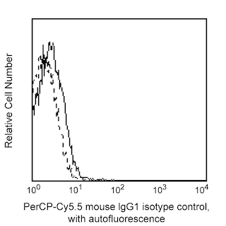
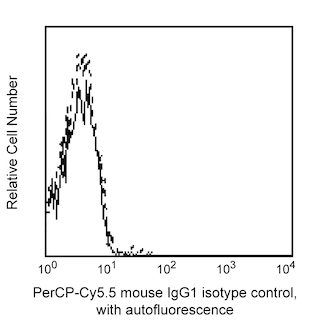
- An isotype control should be used at the same concentration as the antibody of interest.
- Please observe the following precautions: Absorption of visible light can significantly alter the energy transfer occurring in any tandem fluorochrome conjugate; therefore, we recommend that special precautions be taken (such as wrapping vials, tubes, or racks in aluminum foil) to prevent exposure of conjugated reagents, including cells stained with those reagents, to room illumination.
- Cy is a trademark of Amersham Biosciences Limited. This conjugated product is sold under license to the following patents: US Patent Nos. 5,486,616; 5,569,587; 5,569,766; 5,627,027.
- This product is subject to proprietary rights of Amersham Biosciences Corp. and Carnegie Mellon University and made and sold under license from Amersham Biosciences Corp. This product is licensed for sale only for research. It is not licensed for any other use. If you require a commercial license to use this product and do not have one return this material, unopened to BD Biosciences, 10975 Torreyana Rd, San Diego, CA 92121 and any money paid for the material will be refunded.
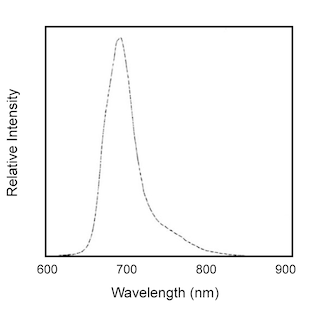
- PerCP-Cy5.5 is optimized for use with a single argon ion laser emitting 488-nm light. Because of the broad absorption spectrum of the tandem fluorochrome, extra care must be taken when using dual-laser cytometers, which may directly excite both PerCP and Cy5.5™. We recommend the use of cross-beam compensation during data acquisition or software compensation during data analysis.
- PerCP-Cy5.5–labelled antibodies can be used with FITC- and R-PE–labelled reagents in single-laser flow cytometers with no significant spectral overlap of PerCP-Cy5.5, FITC, and R-PE fluorescence.
- Source of all serum proteins is from USDA inspected abattoirs located in the United States.
- Caution: Sodium azide yields highly toxic hydrazoic acid under acidic conditions. Dilute azide compounds in running water before discarding to avoid accumulation of potentially explosive deposits in plumbing.
- For fluorochrome spectra and suitable instrument settings, please refer to our Multicolor Flow Cytometry web page at www.bdbiosciences.com/colors.
- Please refer to www.bdbiosciences.com/us/s/resources for technical protocols.
Companion Products


.png?imwidth=320)
The 1G1 monoclonal antibody specifically binds to CD194, also known as the human CC Chemokine Receptor type 4 (CCR4). CCR4 is expressed on activated Th2 cells, regulatory T cells, activated NK cells, basophils, monocytes and platelets. CCR4 is a seven-transmembrane, G-protein-coupled receptor, and is the specific receptor for CC chemokines, CCL22/MDC/Macrophage-Derived Chemokine and CCL17/TARC/Thymus and Activation-Regulated Chemokine. It has been reported that CCR4 mRNA is expressed mainly in the thymus and spleen. The human CCR4 gene has been mapped to chromosome 3p24. The purified form of this antibody has been reported not to be a neutralizing antibody. The immunogen used to generate the 1G1 hybridoma has been reported to be human CCR4 transfected L1.2 mouse lymphoma cells.

Development References (9)
-
Andrew DP, Ruffing N, Kim CH, et al. C-C chemokine receptor 4 expression defines a major subset of circulating nonintestinal memory T cells of both Th1 and Th2 potential. J Immunol. 2000; 166(1):103-111. (Biology). View Reference
-
Bonecchi R, Bianchi G, Bordignon PP, et al. Differential expression of chemokine receptors and chemotactic responsiveness of type 1 T helper cells (Th1s) and Th2s. J Exp Med. 1998; 187(1):129-134. (Biology). View Reference
-
Campbell JJ, Haraldsen G, Pan J, et al. The chemokine receptor CCR4 in vascular recognition by cutaneous but not intestinal memory T cells. Nature. 1999; 400(6746):776-780. (Biology). View Reference
-
D'Ambrosio D, Iellem A, Bonecchi R, et al. Selective up-regulation of chemokine receptors CCR4 and CCR8 upon activation of polarized human type 2 Th cells.. J Immunol. 1998; 161(10):5111-5. (Biology). View Reference
-
Imai T, Baba M, Nishimura M, Kakizaki M, Takagi S, Yoshie O. The T cell-directed CC chemokine TARC is a highly specific biological ligand for CC chemokine receptor 4. J Biol Chem. 1997; 272(23):15036-15042. (Biology). View Reference
-
Imai T, Chantry D, Raport CJ, et al. Macrophage-derived chemokine is a functional ligand for the CC chemokine receptor 4. J Biol Chem. 1998; 273(3):1764-1768. (Biology). View Reference
-
Power CA, Meyer A, Nemeth K, et al. Molecular cloning and functional expression of a novel CC chemokine receptor cDNA from a human basophilic cell line. J Biol Chem. 1995; 270(33):19495-19500. (Biology). View Reference
-
Sallusto F, Lenig D, Mackay CR, Lanzavecchia A. Flexible programs of chemokine receptor expression on human polarized T helper 1 and 2 lymphocytes. J Exp Med. 1998; 187(6):875-883. (Biology). View Reference
-
Samson M, Soularue P, Vassart G, Parmentier M. The genes encoding the human CC-chemokine receptors CC-CKR1 to CC-CKR5 (CMKBR1-CMKBR5) are clustered in the p21.3-p24 region of chromosome 3. Genomics. 1996; 36(3):522-526. (Biology). View Reference
Please refer to Support Documents for Quality Certificates
Global - Refer to manufacturer's instructions for use and related User Manuals and Technical data sheets before using this products as described
Comparisons, where applicable, are made against older BD Technology, manual methods or are general performance claims. Comparisons are not made against non-BD technologies, unless otherwise noted.
For Research Use Only. Not for use in diagnostic or therapeutic procedures.