

Preparation And Storage
Recommended Assay Procedures
Cell Preparation and Staining Procedures for Purified Anti-Human FoxP3 Antibody
1. Bring the buffers to room temperature (RT) before use. Prepare working solutions of the BD Pharmingen Human FoxP3 Buffer Set
Cat. No. 560098 (For the buffer A&C preparations, please see TDS of Cat. No. 560098 buffer instructions for details).
2. Prepare human PBMC. Calculate needed cells per test, (1.2 million) PBMC.
3. Fix cells in bulk or test size using 2 ml of 1x working solution Human FoxP3 Buffer A per test, incubate for
10 min. at RT, protected from light.
Note: Fixed PBMC in bulk may be frozen at -80°C for 24 hours before proceeding to step 4.
4. Wash with 2 ml of BD Pharmingen Stain Buffer (FBS)* per test. Centrifuge 500 x g for 10 minutes and remove
wash buffer.
5. To permeabilize cells in bulk or test size, add 0.5 ml per test of 1x working solution Human FoxP3 Buffer C per test,
incubate for 30 minutes, protected from light.
6. Add an additional 2 ml of BD Pharmingen Stain Buffer (FBS)* per test. Centrifuge 500 x g for 10 minutes
and remove wash buffer.
7. Re-suspend the cells in BD Pharmingen Stain Buffer (FBS)* to 100µl/test; aliquot 100µl of cell suspension per
12 x 75 mm tube.
8. Add purified anti-human FoxP3 mAb at appropriate concentrations at 20µl/test into the tubes. Gently shake or
vortex. Incubate for 30 minutes at RT, protected from light.
9. Wash the cells twice by adding 2 ml of BD Pharmingen Stain Buffer (FBS)* to each tube and centrifuge 500 x g for
5 minutes at RT. Remove wash buffer.
10. Add secondary antibody (APC Rat Anti-Mouse IgG1, Cat. No. 550874) at appropriate concentration. Incubate for 30 minutes at RT protected from light.
11. Repeat wash as in step 9.
12. Block secondary with normal mouse serum 1:10 in 1x PBS, 100 µl per test. Incubate for 10 minutes at RT.
13. Add test volumes of anti-human surface mAbs, incubate for 20 minutes at RT, protected from light.
14. Add 2 ml of BD Pharmingen Stain Buffer (FBS)* to each tube and centrifuge 500 x g for 5 minutes at RT and remove wash buffer.
15. Re-suspend in wash buffer and analyze immediately.
Optional: Add 300µl of 1% formaldehyde in 1x PBS and store at 4°C. Analyze cells within 24 hours.
Note: Caution: Be aware the pellet is buoyant post fixation and careful attention is required to avoid aspiration of the cells.
* We recommend using the BD Pharmingen Stain Buffer (FBS; Cat No. 554656) for initial surface staining and all wash steps and covering tubes during incubation steps with caps or parafilm. We also recommend optimizing forward scatter and side scatter voltages to visualize lymphocytes as separate from debris, red cell ghosts and/or platelets before acquisition.
** Acquire at least 15,000 to 25,000 CD4 positive lymphocytes.
Product Notices
- Since applications vary, each investigator should titrate the reagent to obtain optimal results.
- Caution: Sodium azide yields highly toxic hydrazoic acid under acidic conditions. Dilute azide compounds in running water before discarding to avoid accumulation of potentially explosive deposits in plumbing.
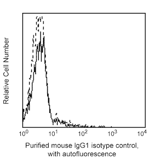
- An isotype control should be used at the same concentration as the antibody of interest.
- Please refer to www.bdbiosciences.com/us/s/resources for technical protocols.
Companion Products

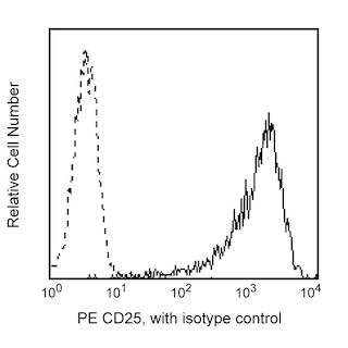
The 259D/C7 monoclonal antibody specifically recognizes human Forkhead box protein P3 (FoxP3) that is also known as Scurfin. FoxP3 is encoded by FOXP3 (Forkhead box P3), likewise known as IPEX (Immune Dysregulation, Polyendocrinopathy, Enteropathy, X-Linked) and JM2, that belongs to the forkhead/winged-helix family of transcriptional regulators. FoxP3 is expressed in CD4+ regulatory T cells (Treg) and represents a specific marker for these cells. Flow cytometric analyses have shown that FoxP3 is expressed by the majority of CD4+CD25+high T cells in peripheral blood while less than half of the CD4+CD25+intermediate cell population are FoxP3 positive. Approximately 5-10% of peripheral CD4+ cells are CD4+CD25+ T regulatory cells. T regulatory cells are thought to play a critical role in the regulation of T cell-mediated immunity and to protect against autoimmunity by suppressing the proliferation and cytokine production of other T cells. In support of this hypothesis, it has been found that Foxp3 is mutated in Scurfy (sf) mice that have defective T cell tolerance leading to an X-linked lymphoproliferative disease. The 259D/C7 antibody recognizes all currently-identified isoforms of human FoxP3 and is crossreactive with FoxP3 from Cynomolgus, Rhesus and Baboon primates.
Development References (3)
-
Brunkow ME, Jeffery EW, Hjerrild KA, et al. Disruption of a new forkhead/winged-helix protein, scurfin, results in the fatal lymphoproliferative disorder of the scurfy mouse. Nat Genet. 2001; 27(1):68-73. (Biology). View Reference
-
Giovanna Roncador et al. Analysis of Foxp3 protein expression in human CD4+CD25+ regulatory Tcells at a single cell level. Eur J Immunol. 2005; 35(Immunogen).
-
Wildin RS, Ramsdell F, Peake J, et al. X-linked neonatal diabetes mellitus, enteropathy and endocrinopathy syndrome is the human equivalent of mouse scurfy. Nat Genet. 2001; 27(1):18-20. (Biology). View Reference
Please refer to Support Documents for Quality Certificates
Global - Refer to manufacturer's instructions for use and related User Manuals and Technical data sheets before using this products as described
Comparisons, where applicable, are made against older BD Technology, manual methods or are general performance claims. Comparisons are not made against non-BD technologies, unless otherwise noted.
For Research Use Only. Not for use in diagnostic or therapeutic procedures.