


Preparation And Storage
Recommended Assay Procedures
For Bioimaging
• Staurosporine (from Streptomyces staurosporeus) is a relatively non-selective protein kinase inhibitor that is often used as a general agent for inducing apoptosis. A stock solution of 2 μM staurosporine should be prepared in culture medium before use.
• This antibody stains HeLa (ATCC CCL-2), A549 (ATCC CCL-185) and U-2 OS (ATCC HTB-96) cells, and it works with either 90% cold methanol or BD Perm/Wash™ buffer permeabilization.
1. Seed the cells in appropriate culture medium at ~10,000 cells per well in a BD Falcon™ 96-well Imaging Plate (Cat. No. 353219), and culture overnight.
2. Add the appropriate concentrations of staurosporine, diluted in 100 μl of culture medium, to the wells and culture for 4 hours.
3. Fix the cells by adding 100 µl of a pre-warmed (37°C) 12% formaldehyde solution to each well and incubating for 30 minutes at room temperature (RT). The final volume is 300 μl per well with a 4% formaldehyde final concentration.
4. Remove the fixative from the wells, and wash the cells twice with 1× PBS.
5. Permeabilize the cells using either -20°C 90% cold methanol (eg. BD Phosflow™ Perm Buffer III, Cat. No. 558050) or BD Perm/Wash™ buffer
(Cat. No. 554723):
a. Add 100 µl of -20°C 90% methanol, BD Phosflow™ Perm Buffer III (Cat. No. 558050), to each well and incubate for 5 minutes.
or b. Add 100 µl of 1× BD Perm/Wash™ buffer to each well and incubate for 30 minutes at RT.
6. Remove the permeabilizer, and wash the wells twice with 100 μl of 1× PBS.
7. Dilute the Alexa Fluor® 647 Mouse anti-Cleaved PARP (Asp 214) antibody conjugate:
a. If the cells were permeabilized with cold methanol, dilute the antibody 1:10 in 1× PBS
or b. If the cells were permeabilized with BD Perm/Wash™ buffer, dilute the antibody 1:10 in 1× BD Perm/Wash™ buffer.
8. Stain the cells by adding 50 µl of the diluted antibody conjugate to each well and incubating for 1 hour at RT or overnight at 4°C, protected
from light.
9. Remove the diluted antibody conjugate, and wash the wells twice with 100 μl of 1X PBS. Remove the PBS.
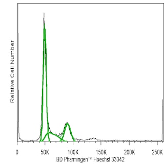
10. To counter-stain the nuclei, dilute Hoechst 33342 Solution (Cat. No. 561908) to 2 μg/ml in 1× PBS, and add 200 μl to each well at least 15 minutes before imaging.
11. View and analyze the cells on an appropriate imaging instrument. Recommended filters for the BD Pathway™ instruments are:
Instrument Excitation Emission Dichroic
BD Pathway 855 620/60 700/75 660 LP
BD Pathway 435 628/40 690/40 FF660
For flow cytometry
• Camptothecin, an extract of the Chinese tree Camptotheca acuminata, has been reported to induce apoptosis in a dose-dependent manner in vitro. A stock solution of 1.0 mM camptothecin (eg, Sigma-Aldrich; Cat. No. C-9911) should be prepared in DMSO before use.
1. Add camptothecin at 4-6 µM final concentration per 1 x 10^6 proliferating Jurkat cells (Human T-cell leukemia; ATCC TIB-152). A control aliquot of untreated cells should also be prepared. Incubate the cells for 4 - 18 hours in a 37°C incubator.
2. Wash the cells twice with cold PBS, resuspend them in BD Cytofix/Cytoperm™ solution at 2 x 10^6 cells/ml, and incubate for 20 minutes on ice.
3. Pellet the cells, and aspirate and discard the BD Cytofix/Cytoperm™ solution.
4. Wash the cells twice at room temperature with 0.5 ml BD Perm/Wash™ buffer per 1 x 10^6 cells, and discard the supernatants.
5. Resuspend the cells in BD Perm/Wash™ buffer at 10 x 10^6 cells/ml, and aliquot test samples of 1 x 10^6 cells per 100-µl test.
6. Add 5 µl Alexa Fluor® 647 Mouse Anti-Cleaved PARP (Asp 214) antibody per test, and incubate for 30 minutes at room temperature, protected from light.
7. Wash each test in 1.0 ml BD Perm/Wash™ Buffer and discard the supernatant.
8. Resuspend each test in 0.5 ml BD Perm/Wash™ Buffer and analyze by flow cytometry.
Product Notices
- This antibody has been developed and certified for the bioimaging application. However, a routine bioimaging test is not performed on every lot. Researchers are encouraged to titrate the reagent for optimal performance.
- This reagent has been pre-diluted for use at the recommended Volume per Test. We typically use 1 × 10^6 cells in a 100-µl experimental sample (a test).
- Alexa Fluor® 647 fluorochrome emission is collected at the same instrument settings as for allophycocyanin (APC).
- For fluorochrome spectra and suitable instrument settings, please refer to our Multicolor Flow Cytometry web page at www.bdbiosciences.com/colors.
- The Alexa Fluor®, Pacific Blue™, and Cascade Blue® dye antibody conjugates in this product are sold under license from Molecular Probes, Inc. for research use only, excluding use in combination with microarrays, or as analyte specific reagents. The Alexa Fluor® dyes (except for Alexa Fluor® 430), Pacific Blue™ dye, and Cascade Blue® dye are covered by pending and issued patents.
- Caution: Sodium azide yields highly toxic hydrazoic acid under acidic conditions. Dilute azide compounds in running water before discarding to avoid accumulation of potentially explosive deposits in plumbing.
- Source of all serum proteins is from USDA inspected abattoirs located in the United States.
- Alexa Fluor® is a registered trademark of Molecular Probes, Inc., Eugene, OR.
- Please refer to www.bdbiosciences.com/us/s/resources for technical protocols.
Companion Products


PARP (Poly [ADP-Ribose] Polymerase) is a 113-kDa nuclear chromatin-associated enzyme that catalyzes the transfer of ADP-ribose units from NAD+ to a variety of nuclear proteins including topoisomerases, histones, and PARP itself. The catalytic activity of PARP is increased in cells following DNA damage, and PARP is thought to play an important role in mediating the normal cellular response to DNA damage. Additionally, PARP is a target of the caspase protease activity associated with apoptosis. The PARP protein consists of an N-terminal DNA-binding domain (DBD) and a C-terminal catalytic domain separated by a central automodification domain. During apoptosis, Caspase-3 cleaves PARP at a recognition site (Asp Glu Val Asp Gly) in the DBD to form 24- and 89-kDa fragments. This process separates the DBD (which is mostly in the 24-kDa fragment) from the catalytic domain (in the 89-kDa fragment) of the enzyme, resulting in the loss of normal PARP function. It has been proposed that inactivation of PARP directs DNA-damaged cells to undergo apoptosis rather than necrotic degradation, and the presence of the 89-kDa PARP cleavage fraction is considered to be a marker of apoptosis.
A peptide corresponding to the N-terminus of the cleavage site (Asp 214) of human PARP was used as the immunogen. The F21-852 monoclonal antibody reacts only with the 89-kDa fragment of human PARP-1 that is downstream of the Caspase-3 cleavage site (Asp214) and contains the automodification and catalytic domains. It does not react with intact human PARP-1. Cross-reactivity with other members of the PARP superfamily is unknown. Recognition of cleaved PARP in mouse cells has been demonstrated, and it may also cross-react with a number of other species due to the conserved nature of the molecule.
Development References (5)
-
Amé J-C, Spenlehauer C, de Murcia G. The PARP superfamily. Bioessays. 2004; 26:882-893. (Biology).
-
Boulares AH, Yakovlev AG, Ivanova V, et al. Role of Poly(ADP-ribose) polymerase (PARP) cleavage in apoptosis. Caspase 3-resistant PARP mutant increases rates of apoptosis in transfected cells. J Biol Chem. 1999; 274(33):22932-22940. (Biology).
-
Cherney BW, McBride OW, Chen D, et al. cDNA sequence, protein structure, and chromosomal location of the human gene for poly(ADP-ribose) polymerase. Proc Natl Acad Sci U S A. 1987; 84(23):8370-8374. (Biology). View Reference
-
D'Amours D, Desnoyers S, D'Silva I, Poirier GG. Poly(ADP-ribosyl)ation reactions in the regulation of nucelar functions. Biochem J. 1999; 342:249-268. (Biology).
-
Soldani G, Scovassi AI. Poly(ADP-ribose) polymerase-1 cleavage during apoptosis: an update. Apoptosis. 2002; 7:321-328. (Biology).
Please refer to Support Documents for Quality Certificates
Global - Refer to manufacturer's instructions for use and related User Manuals and Technical data sheets before using this products as described
Comparisons, where applicable, are made against older BD Technology, manual methods or are general performance claims. Comparisons are not made against non-BD technologies, unless otherwise noted.
For Research Use Only. Not for use in diagnostic or therapeutic procedures.