
Preparation And Storage
Recommended Assay Procedures
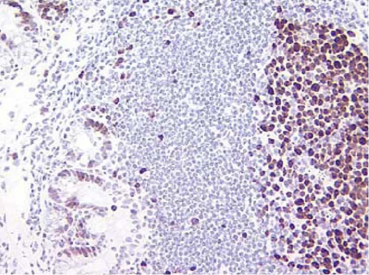
Immunohistochemistry: The B56 clone reactive against Ki-67 is recommended to test for immunohistochemical staining of formalin-fixed paraffin and acetone-fixed frozen sections. For paraffin sections microwave pretreatment with BD Retrievagen A (pH 6.5) (Cat. No. 550524) is required. Tissues tested were human spleen and tonsil. The antibody stains cells in all different stages of proliferation. The isotype control recommended for use with this antibody is purified mouse IgG1 (Cat. No. 550878). For optimal indirect immunohistochemical staining, B56 antibody should be titrated (1:10 to 1:50 dilution) and visualized via a three-step staining procedure in combination with polyclonal, biotin conjugated anti-mouse Igs (multiple adsorbed) (Cat. No. 550337) as the secondary antibody and Streptavidin-HRP (Cat. No. 550946) together with the DAB detection system (Cat. No. 550880).
Product Notices
- Since applications vary, each investigator should titrate the reagent to obtain optimal results.
- An isotype control should be used at the same concentration as the antibody of interest.
- Caution: Sodium azide yields highly toxic hydrazoic acid under acidic conditions. Dilute azide compounds in running water before discarding to avoid accumulation of potentially explosive deposits in plumbing.
- Source of all serum proteins is from USDA inspected abattoirs located in the United States.
- Sodium azide is a reversible inhibitor of oxidative metabolism; therefore, antibody preparations containing this preservative agent must not be used in cell cultures nor injected into animals. Sodium azide may be removed by washing stained cells or plate-bound antibody or dialyzing soluble antibody in sodium azide-free buffer. Since endotoxin may also affect the results of functional studies, we recommend the NA/LE (No Azide/Low Endotoxin) antibody format, if available, for in vitro and in vivo use.
- This antibody has been developed for the immunohistochemistry application. However, a routine immunohistochemistry test is not performed on every lot. Researchers are encouraged to titrate the reagent for optimal performance.
- Species cross-reactivity detected in product development may not have been confirmed on every format and/or application.
- Please refer to www.bdbiosciences.com/us/s/resources for technical protocols.
Companion Products


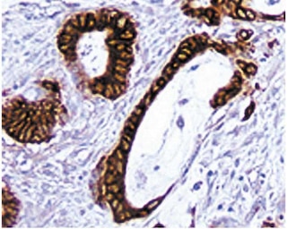
Recognizes Ki-67, a nuclear cell proliferation-associated antigen expressed in all active stages of the cell cycle. Ki-67 is revealed as a double band (345 and 395 kD) in western blot analysis of proliferating cells. B56 was developed using an immunogen composed of the immunodominant epitope of the Ki-67 protein. Antibodies B56 and MIB 1 react with this immunogen. Flow cytometric analysis reveals that the binding of B56-PE can be blocked by MIB 1 purified antibody. Immunohistochemistry analysis demonstrates a staining pattern similar to MIB 1 on both frozen and paraffin-embedded tissue sections.
Development References (14)
-
Benson MJ, Elgueta R, Schpero W, et al. Distinction of the memory B cell response to cognate antigen versus bystander inflammatory signals. J Exp Med. 2009; 206(9):2013-2025. (Clone-specific: Flow cytometry). View Reference
-
Bigley V, Haniffa M, Doulatov S, et al. The human syndrome of dendritic cell, monocyte, B and NK lymphoid deficiency. J Exp Med. 2011; 208(2):227-234. (Clone-specific: Flow cytometry). View Reference
-
Bruno S, Crissman HA, Bauer KD, Darzynkiewicz Z. Changes in cell nuclei during S phase: progressive chromatin condensation and altered expression of the proliferation-associated nuclear proteins Ki-67, cyclin (PCNA), p105, and p34. Exp Cell Res. 1991; 196(1):99-106. (Biology: Flow cytometry). View Reference
-
Bruno S, Darzynkiewicz Z. Cell cycle dependent expression and stability of the nuclear protein detected by Ki-67 antibody in HL-60 cells. Cell Prolif. 1992; 25(1):31-40. (Biology: Flow cytometry). View Reference
-
Kill IR. Localisation of the Ki-67 antigen within the nucleolus: evidence for a fibrillarin-deficient region of the dense fibrillar component. J Cell Sci. 1996; 109(6):1253-1263. (Biology). View Reference
-
Kouro T, Medina KL, Oritani K, Kincade PW. Characteristics of early murine B-lymphocyte precursors and their direct sensitivity to negative regulators. Blood. 2001; 97(9):2708-2715. (Clone-specific: Flow cytometry). View Reference
-
Kubbutat MH, Key G, Duchrow M, Schluter C, Flad HD, Gerdes J. Epitope analysis of antibodies recognising the cell proliferation associated nuclear antigen previously defined by the antibody Ki-67 (Ki-67 protein). J Clin Pathol. 1994; 47(6):524-528. (Biology). View Reference
-
Picker LJ, Hagen SI, Lum R, et al. Insufficient production and tissue delivery of CD4+ memory T cells in rapidly progressive simian immunodeficiency virus infection. J Exp Med. 2004; 200(10):1299-1314. (Clone-specific: Flow cytometry). View Reference
-
Pitcher CJ, Hagen SI, Walker JM, et al. Development and homeostasis of T cell memory in rhesus macaque. J Immunol. 2002; 168(1):29-43. (Clone-specific: Flow cytometry). View Reference
-
Scholzen T, Gerdes J. The Ki-67 protein: from the known and the unknown.. J Cell Physiol. 2000; 182(3):311-22. (Biology). View Reference
-
Shi SR, Key ME, Kalra KL. Antigen retrieval in formalin-fixed, paraffin-embedded tissues: an enhancement method for immunohistochemical staining based on microwave oven heating of tissue sections. J Histochem Cytochem. 1991; 39(6):741-748. (Biology). View Reference
-
Spargo LDJ, Cleland LG, Cockshell MP, Mayrhofer Graham. Recruitment and proliferation of CD4+ T cells in synovium following adoptive transfer of adjuvant-induced arthritis. Int Immunol. 2006; 18(6):897-910. (Clone-specific: Flow cytometry, Immunofluorescence).
-
Starborg M, Gell K, Brundell E, Höög C. The murine Ki-67 cell proliferation antigen accumulates in the nucleolar and heterochromatic regions of interphase cells and at the periphery of the mitotic chromosomes in a process essential for cell cycle progression. J Cell Sci. 1996; 109(1):143-153. (Biology). View Reference
-
Valenti LM, Mathieu J, Chancerelle Y, et al. High levels of endogenous nitric oxide produced after burn injury in rats arrest activated T lymphocytes in the first G1 phase of the cell cycle and then induce their apoptosis. Exp Cell Res. 2005; 306(1):150-167. (Clone-specific: Flow cytometry). View Reference
Please refer to Support Documents for Quality Certificates
Global - Refer to manufacturer's instructions for use and related User Manuals and Technical data sheets before using this products as described
Comparisons, where applicable, are made against older BD Technology, manual methods or are general performance claims. Comparisons are not made against non-BD technologies, unless otherwise noted.
For Research Use Only. Not for use in diagnostic or therapeutic procedures.