Skip to main content Skip to navigation
FITC Rat Anti-Mouse IL-2
Alert icon
This SKU will be discontinuing Apr 2024. Suggested alternate SKU is [554427] or for additional support, contact your local applications specialist. Contact Us #
FITC Rat Anti-Mouse IL-2
Expression of IL-2 by stimulated CD4+ and CD4- BALB/c spleen cells. Splenocytes from 6 month-old BALB/C mice were stimulated for 5 hours with hamster anti-mouse CD3 (2 mg/ml final concentration; Cat. No.553057, clone 145-2C11) and hamster anti-mouse CD28 (2 mg/ml final concentration; Cat. No. 553294, clone 37.51) antibodies in the presence of BD GolgiStop™ (3 µM final concentration; Cat. No. 554724). The cells were harvested, stained with 0.06 µg of PE-conjugated rat anti-mouse CD4 (PE-RM4-5, Cat. No. 553049), fixed, permeabilized, and subsequently stained with 0.06 µg of FITC-conjugated rat anti-mouse IL-2 antibody (FITC-JES6-5H4, Cat. No. 554427) using Pharmingen's staining protocol (left panel). To demonstrate specificity of staining, the binding by FITC-JES6-5H4 was blocked by each of the following: 1) preincubation of the conjugated antibody with recombinant mouse IL-2 (0.12 mg, Cat. No. 550069; middle panel), and by 2) preincubation of the fixed/permeabilized cells with excess unlabelled JES6-5H4 antibody 2.5 mg; Cat. No. 554425; right panel) prior to staining with the FITC-JES6-5H4 antibody. The quadrant markers for the bivariate dot plots were set based on the autofluorescence controls and verified using the recombinant cytokine blocking and unlabelled antibody blocking specificity controls. A suitable rat IgG2b isotype control for assessing the level of background staining on fixed/permeabilized mouse cells is Cat. No. 556923; use at comparable concentrations to antibody of interest.
Product Details
Down Arrow Up Arrow


BD Pharmingen™
Mouse (QC Testing)
Rat IgG2b
Mouse IL-2 Recombinant Protein
Intracellular staining (flow cytometry) (Routinely Tested)
0.5 mg/ml
16183
AB_395385
Aqueous buffered solution containing ≤0.09% sodium azide.
RUO


Preparation And Storage

The monoclonal antibody was purified from tissue culture supernatant or ascites by affinity chromatography. The antibody was conjugated with FITC under optimum conditions, and unreacted FITC was removed. Store undiluted at 4°C and protected from prolonged exposure to light. Do not freeze.

Recommended Assay Procedures

Recommended Assay Procedure:

Immunofluorescent Staining and Flow Cytometric Analysis: The JES6-5H4 antibody is useful for immunofluorescent staining and flow cytometric analysis to identify and enumerate IL-2 producing cells within mixed cell populations. FITC-, PE-, APC, Alexa Fluor® 488 and Alexa Fluor® 647 conjugated antibodies (Cat. No. 554427; Cat. No. 554428; Cat No. 554429, Cat. No. 557725 and Cat. No. 557736) are especially suitable for these studies.  

An appropriate rat IgG2b isotype control is Cat. No. 556923. A useful control for demonstrating specificity of staining is pre-blocking with one of the following: 1) recombinant mouse IL-2 (Cat. No. 550069) or 2) unlabeled JES6-5H4 antibody (Cat. No. 554425), prior to staining. For specific methodology, please visit the protocols section or chapter on intracellular staining in the Immune Function Handbook, both of which are posted on our web site, www.bdbiosciences.com.

Product Notices

  1. Since applications vary, each investigator should titrate the reagent to obtain optimal results.
  2. Please refer to www.bdbiosciences.com/us/s/resources for technical protocols.
  3. Caution: Sodium azide yields highly toxic hydrazoic acid under acidic conditions. Dilute azide compounds in running water before discarding to avoid accumulation of potentially explosive deposits in plumbing.
  4. Use of these products to measure activation antigens expressed on mononuclear cell subsets for the purpose of monitoring immunoregulatory status can fall under one or more claims of the following patents: US Patent Nos. 5,445,939, 5,656,446, 5,843,689; European Patent No. 319,543; Canadian Patent No. 1,296,622; Australian Patent No. 615,880; and Japanese Patent No. 2,769,156.
  5. Alexa Fluor® is a registered trademark of Molecular Probes, Inc., Eugene, OR.
562040 Rev. 1
Antibody Details
Down Arrow Up Arrow
JES6-5H4

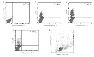
The JES6-5H4 monoclonal antibody specifically binds to mouse interleukin-2 (IL-2), a multifunctional cytokine that plays pivotal roles in immunity and tolerance. It is produced by activated T cells and affects the activation, growth, proliferation and/or differentiation of various cell types including T and B lymphocytes and their precursors, LAK cells, NK cells, and monocytes/macrophages. IL-2 mediates its biological activities by binding to IL-2 receptor complexes. The intermediate affinity IL-2R is comprised of IL-2Rβ (CD122) and common gamma chain (γc; CD132) subunits, whereas the high-affinity IL-2R is comprised of IL-2Rα (CD25), IL-2Rβ, and γc subunits. The JES6-5H4 monoclonal antibody binds to IL-2 and neutralizes its biological activity.

This antibody is routinely tested by flow cytometric analysis. Other applications were tested at BD Biosciences Pharmingen during antibody development only or reported in the literature.

562040 Rev. 1
Format Details
Down Arrow Up Arrow
FITC
Fluorescein (FITC) is part of the BD blue family of dyes. This is a small organic fluorochrome with an excitation maximum (Ex Max) at 494-nm and an emission maximum (Em Max) at 518-nm. FITC is designed to be excited by the Blue laser (488-nm) and detected using an optical filter centered near 520 nm (e.g., a 530/30-nm bandpass filter). Please ensure that your instrument’s configurations (lasers and optical filters) are appropriate for this dye.
altImg
FITC
Blue 488 nm
494 nm
518 nm
562040 Rev.1
Citations & References
Down Arrow Up Arrow

Development References (4)

  1. Abrams J. Immunoenzymetric assay of mouse and human cytokines using NIP-labeled anti-cytokine antibodies. Curr Protoc Immunol. 2001; 1:6.20-6.21. (Clone-specific: ELISA). View Reference
  2. Abrams JS, Roncarolo MG, Yssel H, Andersson U, Gleich GJ, Silver JE. Strategies of anti-cytokine monoclonal antibody development: immunoassay of IL-10 and IL-5 in clinical samples. Immunol Rev. 1992; 127:5-24. (Clone-specific: ELISA, Immunoprecipitation). View Reference
  3. Prussin C, Metcalfe DD. Detection of intracytoplasmic cytokine using flow cytometry and directly conjugated anti-cytokine antibodies. J Immunol Methods. 1995; 188(1):117-128. (Methodology: Flow cytometry). View Reference
  4. Sander B, Hoiden I, Andersson U, Moller E, Abrams JS. Similar frequencies and kinetics of cytokine producing cells in murine peripheral blood and spleen. Cytokine detection by immunoassay and intracellular immunostaining. J Immunol Methods. 1993; 166(2):201-214. (Clone-specific: ELISA). View Reference
View All (4) View Less
562040 Rev. 1

Please refer to Support Documents for Quality Certificates


Global - Refer to manufacturer's instructions for use and related User Manuals and Technical data sheets before using this products as described


Comparisons, where applicable, are made against older BD Technology, manual methods or are general performance claims.  Comparisons are not made against non-BD technologies, unless otherwise noted.

For Research Use Only. Not for use in diagnostic or therapeutic procedures.