Skip to main content Skip to navigation
PerCP-Cy™5.5 Mouse Anti-Human IFN-γ
Alert icon
Consider BD Horizon RealBlue™ 705 (RB705) Reagents, a bright and clean fluorochrome alternative to BD Horizon™ BB700 and PerCP-Cy5.5 More Info #
PerCP-Cy™5.5 Mouse Anti-Human IFN-γ
Flow cytometric analysis for IFN-γ in stimulated human peripheral blood mononuclear cells (PBMC).   Human PBMC were stimulated for 6 hours with 50 ng/mL PMA (Sigma-Aldrich Cat. No. P-8139) and 500 ng/mL calcium ionophore A23187 (Sigma-Aldrich Cat. No. C-9275) in the presence of BD GolgiStop™ (Cat. No. 554724).  Cells were then fixed and permeabilized using BD Cytofix/Cytoperm™ (Cat. No. 554714) followed by staining with either a PerCP-Cy™5.5 Mouse IgG1, κ isotype control (Cat. No. 550795; left panel) or with the  PerCP-Cy™5.5 Mouse Anti-Human IFN-γ antibody (Cat. No. 560704; right panel).  Dot plots were derived from gated events based on light scattering characteristics for lymphocytes.  Flow cytometry was performed on a BD™ LSR II flow cytometry system.  
Product Details
Down Arrow Up Arrow


BD Pharmingen™
IFNG; Interferon-gamma; Interferon-γ; Type II interferon; MAF
Human (QC Testing), Rhesus, Cynomolgus, Baboon (Tested in Development)
Mouse IgG1, κ
Human IFN-γ Recombinant Protein
Intracellular staining (flow cytometry) (Routinely Tested)
5 µl
15978,16176,25712,3458,380793,4049
AB_1727532
Aqueous buffered solution containing BSA and ≤0.09% sodium azide.
RUO


Preparation And Storage

Store undiluted at 4°C and protected from prolonged exposure to light. Do not freeze. The monoclonal antibody was purified from tissue culture supernatant or ascites by affinity chromatography. The antibody was conjugated with PerCP-Cy5.5 under optimum conditions, and unconjugated antibody and free PerCP-Cy5.5 were removed. Storage of PerCP-Cy5.5 conjugates in unoptimized diluent is not recommended and may result in loss of signal intensity.

Recommended Assay Procedures

Flow cytometry:  The B27 antibody is useful for immunofluorescent staining and flow cytometric analysis to identify and enumerate IFN-γ producing cells within mixed cell populations.  A useful control investigators may consider using for demonstrating specificity of staining, is to pre-block with one of the following reagents: (1) recombinant human IFN-γ (Cat. No. 554617) or (2) unlabeled B27 antibody (Cat. No. 554699), prior to staining.

Product Notices

  1. This reagent has been pre-diluted for use at the recommended Volume per Test. We typically use 1 × 10^6 cells in a 100-µl experimental sample (a test).
  2. An isotype control should be used at the same concentration as the antibody of interest.
  3. Source of all serum proteins is from USDA inspected abattoirs located in the United States.
  4. Caution: Sodium azide yields highly toxic hydrazoic acid under acidic conditions. Dilute azide compounds in running water before discarding to avoid accumulation of potentially explosive deposits in plumbing.
  5. Please observe the following precautions: Absorption of visible light can significantly alter the energy transfer occurring in any tandem fluorochrome conjugate; therefore, we recommend that special precautions be taken (such as wrapping vials, tubes, or racks in aluminum foil) to prevent exposure of conjugated reagents, including cells stained with those reagents, to room illumination.
  6. PerCP-Cy5.5 is optimized for use with a single argon ion laser emitting 488-nm light. Because of the broad absorption spectrum of the tandem fluorochrome, extra care must be taken when using dual-laser cytometers, which may directly excite both PerCP and Cy5.5™. We recommend the use of cross-beam compensation during data acquisition or software compensation during data analysis.
  7. PerCP-Cy5.5–labelled antibodies can be used with FITC- and R-PE–labelled reagents in single-laser flow cytometers with no significant spectral overlap of PerCP-Cy5.5, FITC, and R-PE fluorescence.
  8. For fluorochrome spectra and suitable instrument settings, please refer to our Multicolor Flow Cytometry web page at www.bdbiosciences.com/colors.
  9. Species cross-reactivity detected in product development may not have been confirmed on every format and/or application.
  10. Cy is a trademark of GE Healthcare.
  11. Please refer to www.bdbiosciences.com/us/s/resources for technical protocols.
560704 Rev. 2
Antibody Details
Down Arrow Up Arrow
B27

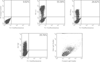
The B27 monoclonal antibody specifically binds to human interferon-γ (IFN-γ), a 14-18 kDa glycoprotein containing 143 amino acid residues.  IFN-γ is a potent multifunctional cytokine produced by several activated cell types including NK, NKT, CD4+TCRαβ+, CD8+TCRαβ+, and TCRγδ+ T cells. IFN-γ exerts its biological effects through specific binding to the high-affinity IFN-γ receptor complex comprised of IFN-γRα (CD119) and IFN-γRβ subunits. In addition to its antiviral effects, IFN-γ upregulates a number of lymphoid cell functions including the antimicrobial and anti-tumor responses of macrophages, NK cells, and neutrophils. In addition, IFN-γ influences the regulation of proliferation, differentiation, and effector responses of B cell and T cell subsets. These influences can involve IFN-γ's capacity to boost MHC class I and II expression by antigen-presenting cells as well as direct effects on B cells and T cells themselves. B27 is a neutralizing antibody. The use of B27 antibody for epitope mapping of human IFN-γ has been described. The B27 antibody has been reported not to bind to denatured IFN-γ.

560704 Rev. 2
Format Details
Down Arrow Up Arrow
PerCP-Cy5.5
PerCP-Cy5.5 dye is part of the BD blue family of dyes. This tandem fluorochrome is comprised of a fluorescent protein complex (PerCP) with an excitation maximum (Ex Max) of 482 nm and an acceptor dye with an emission maximum (Em Max) at 676 nm. PerCP-Cy5 is designed to be excited by the blue laser (488-nm) and detected using an optical filter centered near 680 nm (e.g., a 695/40 nm bandpass filter). The donor dye can be partially excited by the Violet (405-nm) laser resulting in cross-laser excitation and fluorescence spillover. Please ensure that your instrument’s configurations (lasers and optical filters) are appropriate for this dye.
altImg
PerCP-Cy5.5
Blue 488 nm
482 nm
676 nm
560704 Rev.2
Citations & References
Down Arrow Up Arrow

Development References (12)

  1. Czerkinsky C, Andersson G, Ekre HP, Nilsson LA, Klareskog L, Ouchterlony O. Reverse ELISPOT assay for clonal analysis of cytokine production. I. Enumeration of gamma-interferon-secreting cells. J Immunol Methods. 1988; 110(1):29-36. (Biology). View Reference
  2. Farrar MA, Schreiber RD. The molecular cell biology of interferon-gamma and its receptor. Annu Rev Immunol. 1993; 11:571-611. (Biology). View Reference
  3. Favre C, Wijdenes J, Cabrillat H, Djossou O, Banchereau J, de Vries JE. Epitope mapping of recombinant human gamma interferon using monoclonal antibodies. Mol Immunol. 1989; 26(1):17-25. (Biology). View Reference
  4. Fonteneau JF, Le Drean E, Le Guiner S, Gervois N, Diez E, Jotereau F. Heterogeneity of biologic responses of melanoma-specific CTL. J Immunol. 1997; 159(6):2831-2839. (Biology). View Reference
  5. Green JA, Yeh TJ, Overall JC. Rapid, quantitative, semiautomated assay for virus-induced and immune human interferons. J Clin Microbiol. 1980; 12(3):433-438. (Biology). View Reference
  6. Helms T, Boehm BO, Asaad RJ, Trezza RP, Lehmann PV, Tary-Lehmann M. Direct visualization of cytokine-producing recall antigen-specific CD4 memory T cells in healthy individuals and HIV patients. J Immunol. 2000; 164(7):3723-3732. (Biology). View Reference
  7. Meager A, Parti S, Barwick S, Spragg J, O'Hagan K. Detection of hybridomas secreting monoclonal antibodies to human gamma interferon using a rapid screening technique and specificity of certain monoclonal antibodies to gamma interferon. J Interferon Res. 1984; 4(4):619-625. (Biology). View Reference
  8. Meager A. Characterization of interferons and immunoassays. In: Clemens MJ, Morris AG, Gearing AJH, ed. Lymphokines and Interferons. A Practical Approach. Oxford: IRL Press Ltd; 1987:105-127.
  9. Prussin C, Metcalfe DD. Detection of intracytoplasmic cytokine using flow cytometry and directly conjugated anti-cytokine antibodies. J Immunol Methods. 1995; 188(1):117-128. (Methodology: Flow cytometry). View Reference
  10. Rotteveel FT, Kokkelink I, van Lier RA, et al. Clonal analysis of functionally distinct human CD4+ T cell subsets. J Exp Med. 1988; 168(5):1659-1673. (Biology). View Reference
  11. Sester U, Sester M, Hauk M, Kaul H, Köhler H, Girndt M. T-cell activation follows Th1 rather than Th2 pattern in haemodialysis patients.. Nephrol Dial Transplant. 2000; 15(8):1217-1223. (Biology). View Reference
  12. Vogel S, Friedman R, Hogan M. Measurement of antiviral activity induced by interferons a, b, and g.. In: Coligan JE, Kruisbeek AM, Margulies DH, Shevach EM, Strober W, ed. Current Protocols in Immunology. New York: John Wiley & Sons; 2007:6.9.1-6.9.15.
View All (12) View Less
560704 Rev. 2

Please refer to Support Documents for Quality Certificates


Global - Refer to manufacturer's instructions for use and related User Manuals and Technical data sheets before using this products as described


Comparisons, where applicable, are made against older BD Technology, manual methods or are general performance claims.  Comparisons are not made against non-BD technologies, unless otherwise noted.

For Research Use Only. Not for use in diagnostic or therapeutic procedures.