-
Your selected country is
Middle East / Africa
- Change country/language
Old Browser
This page has been recently translated and is available in French now.
Looks like you're visiting us from {countryName}.
Would you like to stay on the current country site or be switched to your country?


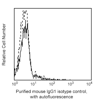
Flow cytometric analysis of CD33 expression on human peripheral blood granulocytes (Left Panel) or monocytes (Right Panel). Human whole blood was stained with either Purified Mouse IgG1 κ Isotype Control (Cat. No. 555746; dashed line histograms) or Purified Mouse Anti-Human CD33 (Cat. No. 555449; solid line histograms). Erythrocytes were lysed with BD Pharm Lyse™ Lysing Buffer (Cat. No. 555899). Fluorescence histograms depicting CD33 (or Ig isotype control) expression were derived from gated events with the side and forward light-scatter characteristics of viable granulocytes or monocytes. Flow cytometry was performed on a BD FACScan™ system.


BD Pharmingen™ Purified Mouse Anti-Human CD33

Regulatory Status Legend
Any use of products other than the permitted use without the express written authorization of Becton, Dickinson and Company is strictly prohibited.
Preparation And Storage
Product Notices
- Since applications vary, each investigator should titrate the reagent to obtain optimal results.
- An isotype control should be used at the same concentration as the antibody of interest.
- Caution: Sodium azide yields highly toxic hydrazoic acid under acidic conditions. Dilute azide compounds in running water before discarding to avoid accumulation of potentially explosive deposits in plumbing.
- Sodium azide is a reversible inhibitor of oxidative metabolism; therefore, antibody preparations containing this preservative agent must not be used in cell cultures nor injected into animals. Sodium azide may be removed by washing stained cells or plate-bound antibody or dialyzing soluble antibody in sodium azide-free buffer. Since endotoxin may also affect the results of functional studies, we recommend the NA/LE (No Azide/Low Endotoxin) antibody format, if available, for in vitro and in vivo use.
- Please refer to http://regdocs.bd.com to access safety data sheets (SDS).
- Please refer to www.bdbiosciences.com/us/s/resources for technical protocols.
Companion Products

.png?imwidth=320)
The WM53 monoclonal antibody specifically recognizes CD33 which is also known as Sialic acid-binding Ig-like lectin 3 (Siglec-3) or gp67. CD33 is a 67 kDa type I transmembrane glycoprotein that is variably expressed on myeloid progenitors, monocytes, macrophages, dendritic cells, neutrophils, basophils, mast cells, and on some activated T cells and NK cells. Normal lymphocytes, platelets, erythrocytes and pluripotent hematopoietic stem cells do not express the CD33 antigen. This glycoprotein reportedly functions as a sialic acid-dependent cell adhesion molecule and this function can be modulated by endogenous sialoglycoconjugates when CD33 is expressed on the membrane.
Development References (6)
-
Favaloro EJ, Bradstock KF, Kabral A, Grimsley P, Zowtyj H, Zola H. Further characterization of human myeloid antigens (gp160,95; gp150; gp67): investigation of epitopic heterogeneity and non-haemopoietic distribution using panels of monoclonal antibodies belonging to CD-11b, CD-13 and CD-33. Br J Haematol. 1988; 69(2):163-171. (Biology). View Reference
-
Favaloro EJ, Moraitis N, Koutts J, Exner T, Bradstock KF. Endothelial cells and normal circulating haemopoietic cells share a number of surface antigens. Thromb Haemost. 1989; 61(2):217-224. (Biology). View Reference
-
Freeman SD, Kelm S, Barber EK, Crocker PR. Characterization of CD33 as a new member of the sialoadhesin family of cellular interaction molecules. Blood. 1995; 85(8):2005-2012. (Biology). View Reference
-
Knapp W. W. Knapp .. et al., ed. Leucocyte typing IV : white cell differentiation antigens. Oxford New York: Oxford University Press; 1989:1-1182.
-
Nakamura Y, Noma M, Kidokoro M, et al. Expression of CD33 antigen on normal human activated T lymphocytes.. Blood. 1994; 83(5):1442-3. (Biology). View Reference
-
van Vugt MJ, van den Herik-Oudijk IE, van de Winkle JG. Binding of PE-CY5 conjugates to the human high-affinity receptor for IgG (CD64). Blood. 1996; 88(6):2358-2361. (Biology). View Reference
Please refer to Support Documents for Quality Certificates
Global - Refer to manufacturer's instructions for use and related User Manuals and Technical data sheets before using this products as described
Comparisons, where applicable, are made against older BD Technology, manual methods or are general performance claims. Comparisons are not made against non-BD technologies, unless otherwise noted.
For Research Use Only. Not for use in diagnostic or therapeutic procedures.
Report a Site Issue
This form is intended to help us improve our website experience. For other support, please visit our Contact Us page.