

Preparation And Storage
Recommended Assay Procedures
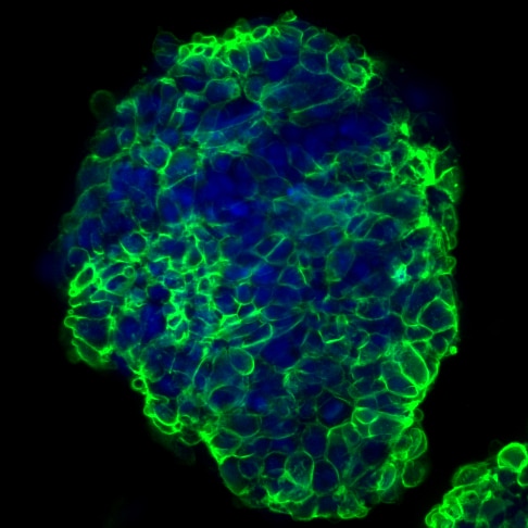
Bioimaging: For more detailed information please refer to "Cellular Imaging" at out website: http://www.bdbiosciences.com/us/s/resources.
BD™ CompBeads can be used as surrogates to assess fluorescence spillover (Compensation). When fluorochrome conjugated antibodies are bound to CompBeads, they have spectral properties very similar to cells. However, for some fluorochromes there can be small differences in spectral emissions compared to cells, resulting in spillover values that differ when compared to biological controls. It is strongly recommended that when using a reagent for the first time, users compare the spillover on cell and CompBead to ensure that BD Comp beads are appropriate for your specific cellular application.
Product Notices
- This reagent has been pre-diluted for use at the recommended Volume per Test. We typically use 1 × 10^6 cells in a 100-µl experimental sample (a test).
- An isotype control should be used at the same concentration as the antibody of interest.
- Caution: Sodium azide yields highly toxic hydrazoic acid under acidic conditions. Dilute azide compounds in running water before discarding to avoid accumulation of potentially explosive deposits in plumbing.
- Source of all serum proteins is from USDA inspected abattoirs located in the United States.
- The Alexa Fluor®, Pacific Blue™, and Cascade Blue® dye antibody conjugates in this product are sold under license from Molecular Probes, Inc. for research use only, excluding use in combination with microarrays, or as analyte specific reagents. The Alexa Fluor® dyes (except for Alexa Fluor® 430), Pacific Blue™ dye, and Cascade Blue® dye are covered by pending and issued patents.
- Alexa Fluor® is a registered trademark of Molecular Probes, Inc., Eugene, OR.
- Alexa Fluor® 647 fluorochrome emission is collected at the same instrument settings as for allophycocyanin (APC).
- For fluorochrome spectra and suitable instrument settings, please refer to our Multicolor Flow Cytometry web page at www.bdbiosciences.com/colors.
- This product is provided under an intellectual property license between Life Technologies Corporation and BD Businesses. The purchase of this product conveys to the buyer the non-transferable right to use the purchased amount of the product and components of the product in research conducted by the buyer (whether the buyer is an academic or for-profit entity). The buyer cannot sell or otherwise transfer (a) this product (b) its components or (c) materials made using this product or its components to a third party or otherwise use this product or its components or materials made using this product or its components for Commercial Purposes. Commercial Purposes means any activity by a party for consideration and may include, but is not limited to: (1) use of the product or its components in manufacturing; (2) use of the product or its components to provide a service, information, or data; (3) use of the product or its components for therapeutic, diagnostic or prophylactic purposes; or (4) resale of the product or its components, whether or not such product or its components are resold for use in research. For information on purchasing a license to this product for any other use, contact Life Technologies Corporation, Cell Analysis Business Unit Business Development, 29851 Willow Creek Road, Eugene, OR 97402, USA, Tel: (541) 465-8300. Fax: (541) 335-0504.
- Please refer to http://regdocs.bd.com to access safety data sheets (SDS).
- Species cross-reactivity detected in product development may not have been confirmed on every format and/or application.
- Accutase is a registered trademark of Innovative Cell Technologies, Inc.
- Please refer to www.bdbiosciences.com/us/s/resources for technical protocols.
The MC480 monoclonal antibody reacts with Stage-Specific Embryonic Antigen-1 (SSEA-1), which is a terminal carbohydrate epitope (3-fucosyl-N-acetyllactosamine or 3-FAL) on glycoproteins and lactose series glycolipids. SSEA-1 is related to Lewis blood group antigens and is found in a variety of embryonic and adult tissues and cancers. As its name implies, the expression of SSEA-1 is stage-specific and can be used to characterize embryonic cells and monitor their differentiation. However, its expression pattern differs in the human and mouse. In the human, SSEA-1 is not found on embryonic stem (ES) cells, embryonic inner cell mass (ICM), or teratocarcinoma (embryonal carcinoma or EC) cells. As human EC and ES cells undergo differentiation, SSEA-1 expression is upregulated. In the adult, the same epitope is expressed as CD15 on granulocytes and monocytes, but not lymphocytes or dendritic cells. In the mouse, SSEA-1 is found on EC, ES, and primordial germ cells, 8-cell to blastocyst embryos, ICM, and on subpopulations of cells in the adult central nervous system, including stem cells. In contrast to the human, SSEA-1 expression is reduced as mouse EC and ES cells undergo differentiation.
Development References (7)
-
Capela A, Temple S. LeX/ssea-1 is expressed by adult mouse CNS stem cells, identifying them as nonependymal. Neuron. 2002; 35:865-875. (Biology).
-
Childs RA, Pennington J, Uemura K, et al. High-molecular-weight glycoproteins are the major carriers of the carbohydrate differentiation antigens I, i and SSEA-1 of mouse teratocarcinoma cells. Biochem J. 1983; 215:491-503. (Clone-specific: Immunofluorescence, Western blot).
-
Draper JS, Pigott C, Thomson JA, Andrews PW. Surface antigens of human embryonic stem cells: changes upon differentiation in culture. J Anat. 2002; 200:249-258. (Clone-specific: Flow cytometry). View Reference
-
Henderson JK, Draper JS, Baillie HS, et al. Preimplantation human embryos and embryonic stem cells show comparable expression of stage-specific embryonic antigens. Stem Cells. 2002; 20:329-337. (Clone-specific: Flow cytometry, Immunofluorescence). View Reference
-
Kannagi R, Nudelman E, Levery SB, Hakomori S. A series of human erythrocyte glycosphingolipids reacting to the monoclonal antibody directed to a developmentally regulated antigen, SSEA-1. J Biol Chem. 1982; 257(24):14865-14874. (Clone-specific).
-
Solter D, Knowles BB. Monoclonal antibody defining a stage-specific mouse embryonic antigen (SSEA-1). Proc Natl Acad Sci U S A. 1978; 75(11):5565-5569. (Immunogen: Cytotoxicity, Radioimmunoassay). View Reference
-
Thomson JA, Itskovitz-Eldor J, Shapiro SS, et al. Embryonic stem cell lines derived from human blastocysts. Science. 1998; 282:1145-1147. (Clone-specific: Immunocytochemistry (cytospins)). View Reference
Please refer to Support Documents for Quality Certificates
Global - Refer to manufacturer's instructions for use and related User Manuals and Technical data sheets before using this products as described
Comparisons, where applicable, are made against older BD Technology, manual methods or are general performance claims. Comparisons are not made against non-BD technologies, unless otherwise noted.
For Research Use Only. Not for use in diagnostic or therapeutic procedures.