

Preparation And Storage
Recommended Assay Procedures
Precautions for flow cytometry: For flow cytometry of leukocytes, it is recommended that Mouse BD Fc Block™ (Cat. No. 553141) be used. When using Mouse BD Fc Block™ (clone 2.4G2), it is important that the second-step anti-hamster IgG antibody does not cross-react with the 2.4G2 mAb (rat IgG2b, κ); we have found that the anti-hamster IgG cocktail (Cat. No. 554010) to be effective. Since this antigen is expressed at low density, it may be desirable to amplify staining by using a biotinylated second-step reagent followed by a "bright" third-step reagent, such as streptavidin-PE (Cat. No. 554061).
Caution: Sodium azide is a reversible inhibitor of oxidative metabolism; therefore, antibody preparations containing this preservative agent must not be used in cell cultures nor injected into animals. Sodium azide may be removed by washing stained cells or plate-bound antibody or dialyzing soluble antibody in sodium azide-free buffer. Since endotoxin may also effect the results of functional studies, we recommend the NA/LE™ (No Azide/Low Endotoxin) antibody format for in vitro and in vivo use.
Product Notices
- Since applications vary, each investigator should titrate the reagent to obtain optimal results.
- Please refer to www.bdbiosciences.com/us/s/resources for technical protocols.
- Although hamster immunoglobulin isotypes have not been well defined, BD Biosciences Pharmingen has grouped Armenian and Syrian hamster IgG monoclonal antibodies according to their reactivity with a panel of mouse anti-hamster IgG mAbs. A table of the hamster IgG groups, Reactivity of Mouse Anti-Hamster Ig mAbs, may be viewed at http://www.bdbiosciences.com/documents/hamster_chart_11x17.pdf.
- Caution: Sodium azide yields highly toxic hydrazoic acid under acidic conditions. Dilute azide compounds in running water before discarding to avoid accumulation of potentially explosive deposits in plumbing.
Companion Products


.png?imwidth=320)
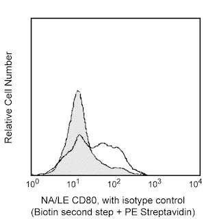
The 16-10A1 monoclonal antibody specifically recognizes CD80 (B7-1). This member of the Ig superfamily, like CD86 (B7-2), can bind to either CD28 or CD152 (CTLA-4) and provide either costimulatory or coinhibitory signals to T cells, respectively. CD80 is constitutively expressed on dendritic cells, monocytes, and peritoneal macrophages as well as by activated B cells and T cells. The 16-10A1 antibody blocks binding of CTLA-4 Ig to CD80 as well as T-cell activation by Con A-elicited peritoneal exudate cells or CD80-transfected cell lines. However, the 16-10A1 antibody alone is not able to block T-cell activation by antigen-presenting cells. The 16-10A1 antibody may reportedly block the binding of another CD80-specific antibody, clone 1G10. In addition, the 16-10A1 antibody may crossreact with an activation antigen expressed on IFN-γ-activated alveolar macrophages of the dog.
This antibody is routinely tested by flow cytometric analysis. Other applications were tested at BD Biosciences Pharmingen during antibody development only or reported in the literature.
Development References (6)
-
Bluestone JA. New perspectives of CD28-B7-mediated T cell costimulation. Immunity. 1995; 2(6):555-559. (Biology). View Reference
-
Boussiotis VA, Gribben JG, Freeman GJ, Nadler LM. Blockade of the CD28 co-stimulatory pathway: a means to induce tolerance. Curr Opin Immunol. 1994; 6(5):797-807. (Biology). View Reference
-
Harlan DM, Hengartner H, Huang ML, et al. Mice expressing both B7-1 and viral glycoprotein on pancreatic beta cells along with glycoprotein-specific transgenic T cells develop diabetes due to a breakdown of T-lymphocyte unresponsiveness. Proc Natl Acad Sci U S A. 1994; 91(8):3137-3141. (Biology: Immunohistochemistry). View Reference
-
Hathcock KS, Laszlo G, Pucillo C, Linsley P, Hodes RJ. Comparative analysis of B7-1 and B7-2 costimulatory ligands: expression and function. J Exp Med. 1994; 180(2):631-640. (Biology). View Reference
-
Herold KC, Vezys V, Koons A, Lenschow D, Thompson C, Bluestone JA. CD28/B7 costimulation regulates autoimmune diabetes induced with multiple low doses of streptozotocin. J Immunol. 1997; 158(2):984-991. (Biology: Immunohistochemistry, In vivo exacerbation). View Reference
-
Razi-Wolf Z, Freeman GJ, Galvin F, Benacerraf B, Nadler L, Reiser H. Expression and function of the murine B7 antigen, the major costimulatory molecule expressed by peritoneal exudate cells. Proc Natl Acad Sci U S A. 1992; 89(9):4210-4214. (Immunogen: Blocking, Immunoprecipitation). View Reference
Please refer to Support Documents for Quality Certificates
Global - Refer to manufacturer's instructions for use and related User Manuals and Technical data sheets before using this products as described
Comparisons, where applicable, are made against older BD Technology, manual methods or are general performance claims. Comparisons are not made against non-BD technologies, unless otherwise noted.
For Research Use Only. Not for use in diagnostic or therapeutic procedures.