
Preparation And Storage
Product Notices
- Since applications vary, each investigator should titrate the reagent to obtain optimal results.
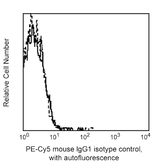
- This antibody has been optimized and preassayed with its matched isotype control to be used at the recommended volume of 20 ul/test. Titration of the reagents or substituting with other (non-matched) isotype control is NOT recommended.
- An isotype control should be used at the same concentration as the antibody of interest.
- Source of all serum proteins is from USDA inspected abattoirs located in the United States.
- Caution: Sodium azide yields highly toxic hydrazoic acid under acidic conditions. Dilute azide compounds in running water before discarding to avoid accumulation of potentially explosive deposits in plumbing.
- For fluorochrome spectra and suitable instrument settings, please refer to our Multicolor Flow Cytometry web page at www.bdbiosciences.com/colors.
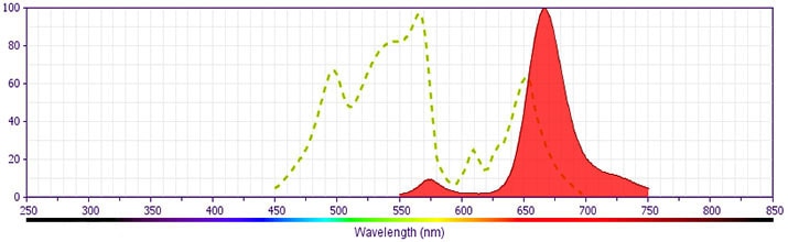
- PE-Cy5 is optimized for use with a single argon ion laser emitting 488-nm light. Because of the broad absorption spectrum of the PE-Cy5 tandem fluorochrome, extra care must be taken when using dual-laser cytometers which may directly excite both PE and Cy5™.
- PE-Cy5 is a tandem fluorochrome composed of R-phycoerythrin (PE), which is excited by the 488 nm light of an Argon ion laser and serves as an energy donor, coupled to the cyanine dye Cy5, which acts as an energy acceptor and fluoresces at 670 nm. BD Biosciences Pharmingen has maximized the fluorochrome energy transfer in PE-Cy5, thus maximizing its fluorescence emission intensity, minimizing residual emission from PE, and minimizing lot-to-lot variation.
- PE-Cy5 tandem fluorochromes have been reported to bind some classes of human macrophages and granulocytes via Fc receptors, and PE has been reported to bind to mouse B lymphocytes via Fc receptors. Preincubation of mouse leukocytes with Mouse BD Fc Block™ purified anti-mouse CD16/CD32 mAb 2.4G2 can reduce the non-specific binding of PE-Cy5-conjugated reagents to mouse B cells. However, PE-Cy5 conjugated reagents should not be used to stain splenocytes of SJL, NOD, and MRL mice as B lymphocytes and/or other leukocytes have been reported to non-specifically stain regardless of the use of Mouse BD Fc Block™ (the CD72c complex has been implicated for PE-Cy5 binding in these strains). Reagents conjugated to PE, PerCP, PerCP-Cy5.5, APC, and APC-Cy7 tandem fluorochrome can be used on leukocytes from these mouse strains.
- Cy is a trademark of GE Healthcare.
- Please observe the following precautions: Absorption of visible light can significantly alter the energy transfer occurring in any tandem fluorochrome conjugate; therefore, we recommend that special precautions be taken (such as wrapping vials, tubes, or racks in aluminum foil) to prevent exposure of conjugated reagents, including cells stained with those reagents, to room illumination.
- Please refer to www.bdbiosciences.com/us/s/resources for technical protocols.
The 9F5 monoclonal antibody specifically binds to CD123. CD123 is the 70 kDa IL-3 receptor α chain (IL-3Rα) that associates with the 120-140 kDa β subunit (CD131/Common β-chain/βc) to form the functional IL-3 receptor complex. The βc chain is also shared with distinct α chain subunits to form the functional heterodimeric receptors for interleukins IL-5 and GM-CSF. IL-3Rα is expressed on a subset of peripheral blood dendritic cells, myeloid precursors, basophils, mast cells, macrophages, and megakaryocytes. Reports indicate that IL-3Rα is also expressed on lymphocytes. The IL-3R plays an important role in hematopoietic progenitor cell growth and differentiation. This antibody does not block binding of IL-3 to the IL-3 receptor.
Development References (2)
-
Macardle PJ, Chen Z, Shih CY, et al. Characterization of human leucocytes bearing the IL-3 receptor. Cell Immunol. 1996; 168(1):59-68. (Biology). View Reference
-
Sun Q, Woodcock JM, Rapoport A, et al. Monoclonal antibody 7G3 recognizes the N-terminal domain of the human interleukin-3 (IL-3) receptor alpha-chain and functions as a specific IL-3 receptor antagonist.. Blood. 1996; 87(1):83-92. (Biology). View Reference
Please refer to Support Documents for Quality Certificates
Global - Refer to manufacturer's instructions for use and related User Manuals and Technical data sheets before using this products as described
Comparisons, where applicable, are made against older BD Technology, manual methods or are general performance claims. Comparisons are not made against non-BD technologies, unless otherwise noted.
For Research Use Only. Not for use in diagnostic or therapeutic procedures.