Skip to main content Skip to navigation
BV421 Mouse Anti-Human CD8b
Alert icon
This product is the replacement for 742390.
BV421 Mouse Anti-Human CD8b
Multiparameter flow cytometric analysis of CD8b expression on Human peripheral blood leucocyte populations. Whole blood was stained with either BD Horizon™ BV421 Mouse IgG2a, k Isotype Control (Cat. No. 562439; Left Plot) or BD Horizon™ BV421 Mouse Anti-Human CD8b antibody (Cat. No. 568373; Right Plot) at 0.25 µg/test. The erythrocytes were lysed with BD FACS™ Lysing Solution (Cat. No. 349202). The bivariate pseudocolor density plot showing the correlated expression of CD8b (or Ig Isotype control staining) versus side-light scatter (SSC-A) signals was derived from gated events with the side and forward light-scattering characteristics of intact leucocyte populations. Flow cytometry and data analysis were performed using a BD LSRFortessa™ X-20 Cell Analyzer System and FlowJo™ software. Data shown on this Technical Data Sheet are not lot specific.
Product Details
Down Arrow Up Arrow


BD Horizon™
CD8B; CD8B1; CD8 beta; CD8β; Leu2; Ly3; LYT3
Human (QC Testing)
Mouse BALB/c IgG2a, κ
CD8+ suppressor T-cell
Flow cytometry (Routinely Tested)
0.2 mg/ml
Aqueous buffered solution containing ≤0.09% sodium azide.
RUO


Preparation And Storage

Store undiluted at 4°C and protected from prolonged exposure to light. Do not freeze. The monoclonal antibody was purified from tissue culture supernatant or ascites by affinity chromatography. The antibody was conjugated to the dye under optimum conditions and unconjugated antibody and free dye were removed.

Recommended Assay Procedures

BD® CompBeads can be used as surrogates to assess fluorescence spillover (compensation).  When fluorochrome conjugated antibodies are bound to BD® CompBeads, they have spectral properties very similar to cells.   However, for some fluorochromes there can be small differences in spectral emissions compared to cells, resulting in spillover values that differ when compared to biological controls.  It is strongly recommended that when using a reagent for the first time, users compare the spillover on cells and BD® CompBeads to ensure that BD® CompBeads are appropriate for your specific cellular application.

For optimal and reproducible results, BD Horizon Brilliant Stain Buffer should be used anytime BD Horizon Brilliant dyes are used in a multicolor flow cytometry panel.  Fluorescent dye interactions may cause staining artifacts which may affect data interpretation.  The BD Horizon Brilliant Stain Buffer was designed to minimize these interactions. When BD Horizon Brilliant Stain Buffer is used in in the multicolor panel, it should also be used in the corresponding compensation controls for all dyes to achieve the most accurate compensation. For the most accurate compensation, compensation controls created with either cells or beads should be exposed to BD Horizon Brilliant Stain Buffer for the same length of time as the corresponding multicolor panel. More information can be found in the Technical Data Sheet of the BD Horizon Brilliant Stain Buffer (Cat. No. 563794/566349) or the BD Horizon Brilliant Stain Buffer Plus (Cat. No. 566385).

Product Notices

  1. Please refer to www.bdbiosciences.com/us/s/resources for technical protocols.
  2. Caution: Sodium azide yields highly toxic hydrazoic acid under acidic conditions. Dilute azide compounds in running water before discarding to avoid accumulation of potentially explosive deposits in plumbing.
  3. For fluorochrome spectra and suitable instrument settings, please refer to our Multicolor Flow Cytometry web page at www.bdbiosciences.com/colors.
  4. Since applications vary, each investigator should titrate the reagent to obtain optimal results.
  5. An isotype control should be used at the same concentration as the antibody of interest.
  6. BD Horizon Brilliant Violet 421 is covered by one or more of the following US patents: 8,158,444; 8,362,193; 8,575,303; 8,354,239.
  7. BD Horizon Brilliant Stain Buffer is covered by one or more of the following US patents: 8,110,673; 8,158,444; 8,575,303; 8,354,239.
  8. Please refer to http://regdocs.bd.com to access safety data sheets (SDS).
  9. Pacific Blue™ is a trademark of Life Technologies Corporation.
  10. Human donor specific background has been observed in relation to the presence of anti-polyethylene glycol (PEG) antibodies, developed as a result of certain vaccines containing PEG, including some COVID-19 vaccines. We recommend use of BD Horizon Brilliant™ Stain Buffer in your experiments to help mitigate potential background. For more information visit https://www.bdbiosciences.com/en-us/support/product-notices.
568373 Rev. 1
Antibody Details
Down Arrow Up Arrow
2ST8.5H7

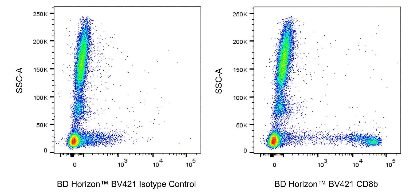
The 2ST8.5H7 monoclonal antibody specifically recognizes an epitope formed by the combination of CD8 alpha and beta chains. The majority of peripheral blood CD8+ T lymphocytes expresses a CD8αβ heterodimer (32, 30 kilodaltons (kDa)), while CD8+CD16+ natural killer (NK) cells and CD8+ TCR γδ+ T lymphocytes express CD8αα homodimers. The 2ST8.5H7 antibody can therefore be used to selectively bind to CD8+ T cells while excluding CD8+ NK cells. CD8 binds to class I major histocompatibility (MHC) molecules, resulting in increased adhesion between the CD8+ T lymphocytes and target cells. Binding of CD8 to class I MHC molecules enhances the activation of resting T lymphocytes. CD8 is coupled to a protein tyrosine kinase, p56lck. The CD8:p56lck complex can play a role in T-lymphocyte activation through mediation of the interactions between CD8 and the CD3/TCR complex. The CD8β antigen is present on the human suppressor/cytotoxic T-lymphocyte subset. The CD8 antigen is expressed on 19% to 48% of normal peripheral blood lymphocytes and 60% to 85% of normal thymocytes. The 2ST8.5H7 antibody crossreacts with lymphocytes of some nonhuman primate species.

568373 Rev. 1
Format Details
Down Arrow Up Arrow
BV421
The BD Horizon Brilliant Violet™ 421 (BV421) Dye is part of the BD Horizon Brilliant Violet™ family of dyes. This polymer-technology based dye has an excitation maximum (Ex Max) of 407-nm and an emission maximum (Em Max) at 423-nm. Driven by BD innovation, BV421 is designed to be excited by the violet laser (405-nm) and detected using an optical filter centered near 420-nm (e.g., a 431/28-nm or 450/50-nm bandpass filter). BV421 is an ideal alternative for V450 as it is approximately ten times brighter with less spillover into the BV510/V500 detector. Please ensure that your instrument’s configurations (lasers and optical filters) are appropriate for this dye.
altImg
BV421
Violet 405 nm
407 nm
423 nm
568373 Rev.1
Citations & References
Down Arrow Up Arrow

Development References (6)

  1. Hambor JE, Weber MC, Tykocinski ML, Kaplan DR. Regulation of allogeneic responses by expression of CD8 alpha chain on stimulator cells.. Int Immunol. 1990; 2(9):879-83. (Clone-specific: Flow cytometry). View Reference
  2. Hori T, Cupp J, Wrighton N, Lee F, Spits H. Identification of a novel human thymocyte subset with a phenotype of CD3- CD4+ CD8 alpha + beta-1. Possible progeny of the CD3- CD4- CD8- subset.. J Immunol. 1991; 146(12):4078-84. (Clone-specific: Flow cytometry). View Reference
  3. Ledbetter JA, Evans RL, Lipinski M, Cunningham-Rundles C, Good RA, Herzenberg LA. Evolutionary conservation of surface molecules that distinguish T lymphocyte helper/inducer and cytotoxic/suppressor subpopulations in mouse and man. J Exp Med. 1981; 153(2):310-323. (Biology). View Reference
  4. Moebius U. Cluster report: CD8. In: Knapp W. W. Knapp .. et al., ed. Leucocyte typing IV : white cell differentiation antigens. Oxford New York: Oxford University Press; 1989:342-343.
  5. Shiue L, Gorman SD, Parnes JR. A second chain of human CD8 is expressed on peripheral blood lymphocytes.. J Exp Med. 1988; 168(6):1993-2005. (Clone-specific: Flow cytometry). View Reference
  6. Terry LA, DiSanto JP, Small TN, Flomenberg N. Differential expression of the CD8 and Lyt-3 antigens on a subset of human T-cell receptor γ/δ-bearing lymphocytes. In: Knapp W. W. Knapp .. et al., ed. Leucocyte typing IV : white cell differentiation antigens. Oxford New York: Oxford University Press; 1989:345-346.
View All (6) View Less
568373 Rev. 1

Please refer to Support Documents for Quality Certificates


Global - Refer to manufacturer's instructions for use and related User Manuals and Technical data sheets before using this products as described


Comparisons, where applicable, are made against older BD Technology, manual methods or are general performance claims.  Comparisons are not made against non-BD technologies, unless otherwise noted.

For Research Use Only. Not for use in diagnostic or therapeutic procedures.