Skip to main content Skip to navigation
BUV805 Rat Anti-CD11b
Alert icon
This product is the replacement for 741934.
BUV805 Rat Anti-CD11b
Multiparameter flow cytometric analysis of CD11b expression on mouse bone-marrow cells. Mouse bone-marrow cells were preincubated with Purified Rat Anti-Mouse CD16/CD32 antibody (Mouse BD Fc Block™) (Cat. No. 553141/553142). The cells were then stained with either BD Horizon™ BUV805 Rat IgG2b, κ Isotype Control (Cat. No. 612901; Left Plot) or BD Horizon™ BUV805 Rat Anti-Mouse CD11b antibody (Cat. No. 568345; Right Plot) at 0.25 µg/test. BD Via-Probe™ Cell Viability 7-AAD Solution (Cat. No. 555815/555816) was added to cells right before analysis. A bivariate pseudocolor density plot showing the correlated expression of CD11b (or Ig Isotype control staining) versus side-light scatter (SSC-A) signals was derived from gated events with the forward and side-light scatter characteristics of viable (7-AAD-negative) bone marrow cell populations. Flow cytometry and data analysis were performed using a BD LSRFortessa™ X-20 Cell Analyzer System and FlowJo™ software. Data shown on this Technical Data Sheet are not lot specific.
BUV805 Rat Anti-CD11b
Multiparameter flow cytometric analysis of CD11b expression on human peripheral blood leucocytes. Whole blood was stained with either BD Horizon™ BUV805 Rat IgG2b, κ Isotype Control (Cat. No. 612901; Left Plot) or BD Horizon™ BUV805 Mouse Anti-Human CD11b antibody (Cat. No. 568345; Right Plot) at 0.25 µg/test. Erythrocytes were lysed with BD FACS™ Lysing Solution (Cat. No. 349202). A bivariate pseudocolor density plot showing the correlated expression of CD11b (or Ig Isotype control staining) versus side-light scatter (SSC-A) signals was derived from gated events with the forward and side-light scatter characteristics of intact leucocyte populations. Flow cytometry and data analysis were performed using a BD LSRFortessa™ X-20 Cell Analyzer System and FlowJo™ software. Data shown on this Technical Data Sheet are not lot specific.
Multiparameter flow cytometric analysis of CD11b expression on mouse bone-marrow cells. Mouse bone-marrow cells were preincubated with Purified Rat Anti-Mouse CD16/CD32 antibody (Mouse BD Fc Block™) (Cat. No. 553141/553142). The cells were then stained with either BD Horizon™ BUV805 Rat IgG2b, κ Isotype Control (Cat. No. 612901; Left Plot) or BD Horizon™ BUV805 Rat Anti-Mouse CD11b antibody (Cat. No. 568345; Right Plot) at 0.25 µg/test. BD Via-Probe™ Cell Viability 7-AAD Solution (Cat. No. 555815/555816) was added to cells right before analysis. A bivariate pseudocolor density plot showing the correlated expression of CD11b (or Ig Isotype control staining) versus side-light scatter (SSC-A) signals was derived from gated events with the forward and side-light scatter characteristics of viable (7-AAD-negative) bone marrow cell populations. Flow cytometry and data analysis were performed using a BD LSRFortessa™ X-20 Cell Analyzer System and FlowJo™ software. Data shown on this Technical Data Sheet are not lot specific.
Multiparameter flow cytometric analysis of CD11b expression on human peripheral blood leucocytes. Whole blood was stained with either BD Horizon™ BUV805 Rat IgG2b, κ Isotype Control (Cat. No. 612901; Left Plot) or BD Horizon™ BUV805 Mouse Anti-Human CD11b antibody (Cat. No. 568345; Right Plot) at 0.25 µg/test. Erythrocytes were lysed with BD FACS™ Lysing Solution (Cat. No. 349202). A bivariate pseudocolor density plot showing the correlated expression of CD11b (or Ig Isotype control staining) versus side-light scatter (SSC-A) signals was derived from gated events with the forward and side-light scatter characteristics of intact leucocyte populations. Flow cytometry and data analysis were performed using a BD LSRFortessa™ X-20 Cell Analyzer System and FlowJo™ software. Data shown on this Technical Data Sheet are not lot specific.
Product Details
Down Arrow Up Arrow


BD Horizon™
Itgam; Integrin alpha-M; Ly-40; Mac-1a; Mac-1 alpha; CR3A; CR-3 alpha chain
Mouse (QC Testing), Human (Tested in Development)
Rat DA, also known as DA/HA IgG2b, κ
Mouse Splenic Cells
Flow cytometry (Routinely Tested)
0.2 mg/ml
16409
Aqueous buffered solution containing ≤0.09% sodium azide.
RUO


Preparation And Storage

Store undiluted at 4°C and protected from prolonged exposure to light. Do not freeze. The monoclonal antibody was purified from tissue culture supernatant or ascites by affinity chromatography. The antibody was conjugated to the dye under optimum conditions and unconjugated antibody and free dye were removed.

Recommended Assay Procedures

   BD® CompBeads can be used as surrogates to assess fluorescence spillover (compensation). When fluorochrome-conjugated antibodies are bound to BD® CompBeads, they have spectral properties very similar to cells. However, for some fluorochromes there can be small differences in spectral emissions compared to cells, resulting in spillover values that differ when compared to biological controls. It is strongly recommended that when using a reagent for the first time, users compare the spillover on cells and BD® CompBeads. This will ensure that BD® CompBeads are appropriate for your specific cellular application.

   For optimal and reproducible results, BD Horizon Brilliant™ Stain Buffer should be used anytime BD Horizon Brilliant dyes are used in a multicolor flow cytometry panel.  Fluorescent dye interactions may cause staining artifacts which may affect data interpretation.  The BD Horizon Brilliant Stain Buffer was designed to minimize these interactions. When BD Horizon Brilliant Stain Buffer is used in in the multicolor panel, it should also be used in the corresponding compensation controls for all dyes to achieve the most accurate compensation. For the most accurate compensation, compensation controls created with either cells or beads should be exposed to BD Horizon Brilliant Stain Buffer for the same length of time as the corresponding multicolor panel. More information can be found in the Technical Data Sheet of the BD Horizon Brilliant Stain Buffer (Cat. No. 563794/566349) or the BD Horizon Brilliant Stain Buffer Plus (Cat. No. 566385).

   Note:  When using high concentrations of antibody, background binding of this dye to erythroid cell subsets (mature erythrocytes and precursors) has been observed.  For researchers studying these cell populations, or in cases where light scatter gating does not adequately exclude these cells from the analysis, this background may be an important factor to consider when selecting reagents for panel(s).

Product Notices

  1. Please refer to www.bdbiosciences.com/us/s/resources for technical protocols.
  2. Caution: Sodium azide yields highly toxic hydrazoic acid under acidic conditions. Dilute azide compounds in running water before discarding to avoid accumulation of potentially explosive deposits in plumbing.
  3. Since applications vary, each investigator should titrate the reagent to obtain optimal results.
  4. For fluorochrome spectra and suitable instrument settings, please refer to our Multicolor Flow Cytometry web page at www.bdbiosciences.com/colors.
  5. An isotype control should be used at the same concentration as the antibody of interest.
  6. Species cross-reactivity detected in product development may not have been confirmed on every format and/or application.
  7. BD Horizon Brilliant Ultraviolet 805 is covered by one or more of the following US patents: 8,110,673, 8,158,444; 8,227,187; 8,575,303; 8,354,239.
  8. BD Horizon Brilliant Stain Buffer is covered by one or more of the following US patents: 8,110,673; 8,158,444; 8,575,303; 8,354,239.
  9. Please refer to http://regdocs.bd.com to access safety data sheets (SDS).
  10. Human donor specific background has been observed in relation to the presence of anti-polyethylene glycol (PEG) antibodies, developed as a result of certain vaccines containing PEG, including some COVID-19 vaccines. We recommend use of BD Horizon Brilliant™ Stain Buffer in your experiments to help mitigate potential background. For more information visit https://www.bdbiosciences.com/en-us/support/product-notices.
568345 Rev. 1
Antibody Details
Down Arrow Up Arrow
M1/70

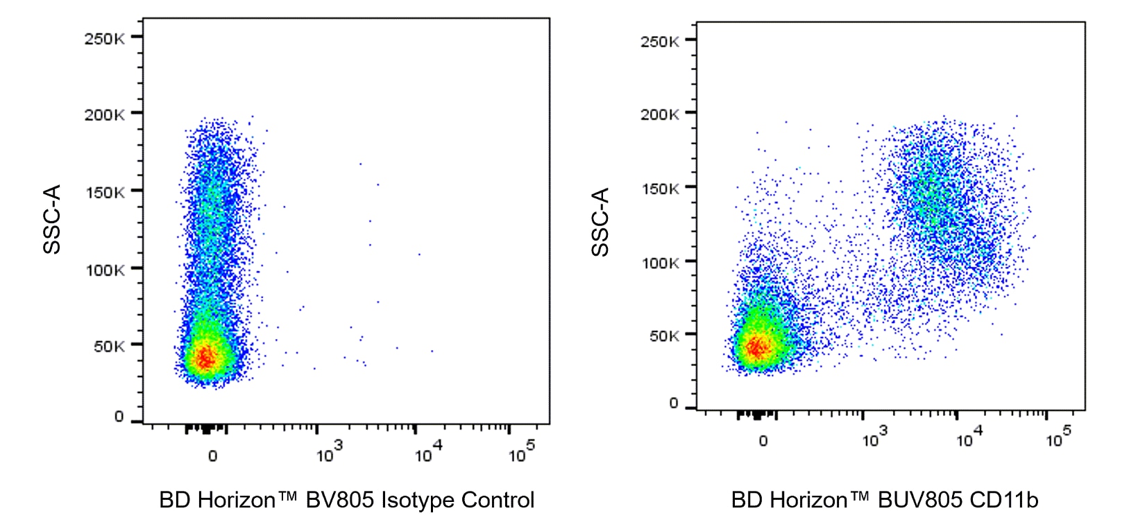
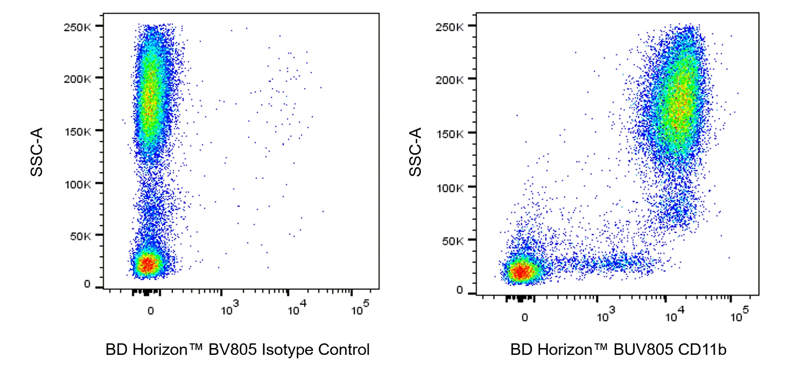
The M1/70 monoclonal antibody specifically binds to CD11b, also known as Integrin alpha M (Itgam or αM). CD11b is a 170-kDa type 1 transmembrane glycoprotein and belongs to the Integrin alpha chain family. CD11b serves as the alpha chain of the heterodimeric Mac-1 integrin (CD11b/CD18, αMβ2), also known as complement receptor 3 (CR3). Mac-1 mediates adhesion to ICAM-1 (CD54), ICAM-2 (CD102), fibrinogen and binding to C3bi.  Mac-1 is expressed at varying levels on granulocytes, macrophages, myeloid-derived dendritic cells, natural killer cells, microglia, and B-1 B lymphocytes.  Mac-1 expression is rapidly upregulated on neutrophils after activation, in the same time period that CD62L (L-selectin) is shed from the cell surface.  The M1/70 antibody reportedly blocks cell adherence and C3bi binding but does not block cell-mediated lysis.  Cross-reaction of the M1/70 antibody with CD11b expressed on human monocytes, polymorphonuclear leukocytes, and NK cells has been reported.

568345 Rev. 1
Format Details
Down Arrow Up Arrow
BUV805
The BD Horizon Brilliant™ Ultraviolet 805 (BUV805) Dye is part of the BD Horizon Brilliant™ Ultraviolet family of dyes. This tandem fluorochrome is comprised of a BUV395 donor with an excitation maximum (Ex Max) of 351-nm and an acceptor dye with an emission maximum (Em Max) at 803-nm. BUV805, driven by BD innovation, is designed to be excited by the ultraviolet laser (355 nm) and detected using an optical filter centered near 805-nm (e.g., a 820/60 or a 780/60 bandpass filter). Please ensure that your instrument’s configurations (lasers and optical filters) are appropriate for this dye.
altImg
BUV805
Ultraviolet 355 nm
351 nm
803 nm
568345 Rev.1
Citations & References
Down Arrow Up Arrow

Development References (12)

  1. Ault KA, Springer TA. Cross-reaction of a rat-anti-mouse phagocyte-specific monoclonal antibody (anti-Mac-1) with human monocytes and natural killer cells. J Immunol. 1981; 126(1):359-364. (Clone-specific: Flow cytometry, Fluorescence activated cell sorting, Radioimmunoassay). View Reference
  2. Beller DI, Springer TA, Schreiber RD. Anti-Mac-1 selectively inhibits the mouse and human type three complement receptor. J Exp Med. 1982; 156(4):1000-1009. (Clone-specific: Blocking, Inhibition). View Reference
  3. Driver DJ, McHeyzer-Williams LJ, Cool M, Stetson DB, McHeyzer-Williams MG. Development and maintenance of a B220- memory B cell compartment. J Immunol. 2001; 167(3):1393-1405. (Clone-specific: Flow cytometry, Immunofluorescence). View Reference
  4. Kaji K, Takeshita S, Miyake K, Takai T, Kudo A. Functional association of CD9 with the Fc gamma receptors in macrophages. J Immunol. 2001; 166(5):3256-3265. (Clone-specific: Fluorescence microscopy, Immunofluorescence). View Reference
  5. Kishimoto TK, Jutila MA, Berg EL, Butcher EC. Neutrophil Mac-1 and MEL-14 adhesion proteins inversely regulated by chemotactic factors. Science. 1989; 245(4923):1238-1241. (Biology). View Reference
  6. Lagasse E, Weissman IL. Flow cytometric identification of murine neutrophils and monocytes. J Immunol Methods. 1996; 197(1-2):139-150. (Clone-specific: Flow cytometry). View Reference
  7. Lub M, van Kooyk Y, Figdor CG. Competition between lymphocyte function-associated antigen 1 (CD11a/CD18) and Mac-1 (CD11b/CD18) for binding to intercellular adhesion molecule-1 (CD54). J Leukoc Biol. 1996; 59(5):648-655. (Clone-specific: Immunoprecipitation). View Reference
  8. Sanchez-Madrid F, Simon P, Thompson S, Springer TA. Mapping of antigenic and functional epitopes on the alpha- and beta-subunits of two related mouse glycoproteins involved in cell interactions, LFA-1 and Mac-1. J Exp Med. 1983; 158(2):586-602. (Clone-specific: Immunoaffinity chromatography, Immunoprecipitation, Inhibition). View Reference
  9. Springer T, Galfre G, Secher D, Milstein C. Monoclonal xenogeneic antibodies to mouse leukocyte antigens: identification of macrophage-specific and other differentiation antigens. Curr Top Microbiol Immunol. 1978; 81:45-50. (Immunogen: Immunoprecipitation, Radioimmunoassay). View Reference
  10. Springer T, Galfre G, Secher DS, Milstein C. Mac-1: a macrophage differentiation antigen identified by monoclonal antibody. Eur J Immunol. 1979; 9(4):301-306. (Clone-specific: Flow cytometry, Immunoprecipitation). View Reference
  11. Springer T, Galfre G, Secher DS, Milstein C. Monoclonal xenogeneic antibodies to murine cell surface antigens: identification of novel leukocyte differentiation antigens. Eur J Immunol. 1978; 8(8):539-551. (Immunogen: Immunoprecipitation). View Reference
  12. Springer TA, Davignon D, Ho MK, Kurzinger K, Martz E, Sanchez-Madrid F. LFA-1 and Lyt-2,3, molecules associated with T lymphocyte-mediated killing; and Mac-1, an LFA-1 homologue associated with complement receptor function. Immunol Rev. 1982; 68:171-195. (Immunogen: Immunoprecipitation, Radioimmunoassay). View Reference
View All (12) View Less
568345 Rev. 1

Please refer to Support Documents for Quality Certificates


Global - Refer to manufacturer's instructions for use and related User Manuals and Technical data sheets before using this products as described


Comparisons, where applicable, are made against older BD Technology, manual methods or are general performance claims.  Comparisons are not made against non-BD technologies, unless otherwise noted.

For Research Use Only. Not for use in diagnostic or therapeutic procedures.