BD Pharmingen™ HumanTh17/Treg Phenotyping Kit
(RUO)
Description
Components:
51-9006636 Human Th17/Treg Phenotyping Cocktail 1.0 ml
51-9005451 Human FoxP3 Buffer A (10 X) 25 ml
51-9005450 Human FoxP3 Buffer B (50 X) 1.0 ml
51-2092KZ BD GolgiStop™ Protein Transport Inhibitor (containing monensin) 0.7 ml
The immune system protects individuals from a broad range of pathogenic microorganisms while avoiding inappropriate or extreme immune responses, such as autoimmune responses, that could be harmful. The peripheral CD4+ T cell pool includes multiple functionally distinct T cell subsets that arise through thymic differentiation or as a consequence of antigen-driven expansion and differentiation of peripheral naïve T cells. The early response of naïve CD4+ T cells to antigenic stimulation may be characterized by high level proliferation and a limited cytokine repertoire. Further differentiation yields cells with a more diverse potential for cytokine expression. Depending upon the balance of local cytokines, costimulatory molecules, antigen levels, and genetic factors, Th17 effector/memory cells or inducible CD4+ T regulatory cells (iTreg) can be generated from the naïve CD4+ T cell pool. In addition, the peripheral T cell pool contains natural CD4+ T regulatory cells (nTreg) that are generated in the thymus as a functionally mature subpopulation of T cells.
Functionally-polarized CD4+ T cell subsets have been identified based on their distinctive patterns of cytokine secretion, transcription factor expression and function. As a signature cytokine, Th17 express high levels of interleukin-17A (IL-17A) whereas Treg are characterized by the expression of the FoxP3 transcription factor. Through the secretion of IL-17A and other factors, Th17 cells recruit and activate neutrophils and mediate immune responses against extracellular bacteria and fungi. Th17 cells are also implicated in mediating autoimmune responses. Natural and inducible Tregs can suppress the function of other T cells or cell types involved in the immune response through a variety of mechanisms including cytokines. In this way, Treg safeguard against the immune system's responsiveness to self antigens and restrain excessive responsiveness to foreign antigens that could be harmful to the host.
The Th17/Treg paradigm provides a useful model system for investigating the cellular and molecular mechanisms that mediate protective as well as harmful immune responses including autoimmune diseases. The BD Human Th17/Treg Phenotyping Kit provides an easy-to-use three-color cocktail of fluorescent antibodies-specific for human CD4, IL-17A (for Th17) and Foxp3 (for Treg)-that will enable researchers to identify and characterize the nature of the CD4+ T cell types present in their system by multicolor flow cytometric analysis. The kit can be used to successfully analyze ex vivo lymphoid cell samples (eg, for the types of in vivo-generated human peripheral blood CD4+ T cells) or to monitor CD4+ T cell differentiation by cells cultured within various experimental model systems.
Investigators should note that the appearance of BD GolgiStop™ Protein Transport Inhibitor may range in color from clear (colorless) to light yellow.
Preparation And Storage
Recommended Assay Procedures
General Protocol for Human Th17/Treg Phenotyping Kit
A. Stimulation of the Cells
Various in vitro methods have been reported for polarization or stimulation of T helper cell subsets of which PMA (Phorbol ester) plus Ionomycin (Calcium Ionophore) has been particularly useful for quickly inducing and characterizing polyclonal cytokine-producing cells. For this kit we recommend the stimulation of normal PBMC in media for 5h with PMA/Ionomycin (at 50 ng/ml and 1μg/ml respectively for 1 million cells) in the presence of BD GolgiStop™ Protein Transport Inhibitor (Cat No. 560762, 51-2092KZ). Add 4 μl of BD GolgiStop™ for every 6 ml of cell culture and mix thoroughly. It is recommended that BD GolgiStop not be kept in cell culture for longer than 12 hours. Note: Kinetic studies need to be performed to determine the optimal incubation time for each experimental system.
B. Staining of the Cells
1. Harvest of the Cells: Collect cells from in vitro stimulatory cultures treated with a protein transport inhibitor. Spin down (250 X g) cells out of the medium containing BD GolgiStop™, and suspend in BD Pharmingen™ Stain Buffer (FBS)*; Cat No. 554656. Count cells and adjust concentration to 20 million cells/ml in Stain Buffer (FBS).
2. Fixing and Staining Procedures for the cocktail
Preparation of Buffers before Use
Note: The working solutions for Human FoxP3 Buffers A and C need to be made fresh for each experimental set.
• Dilute FoxP3 Buffer A (10X concentrate) 1:10 with room temperature (20°C to 25°C) deionized water.
• To make a working solution of Buffer C, dilute FoxP3 Buffer B into 1X FoxP3 Buffer A at a ratio of 1:50
(Buffer B:Buffer A).
a. To fix the cells, gently re-suspend pellet of 1 million cells in residual volume of wash buffer and then add 2ml of 1x Human FoxP3 Buffer A. Vortex. Incubate for 10-20 minutes at RT in the dark.
b. Centrifuge 500 x g for 5 minutes, and remove fixative. Caution: Be aware the pellet is buoyant. Note: The pellet after fixation is loose and care should be taken when aspirating the wash buffer from the tubes. DO NOT ASPIRATE ALL of the buffer but leave around 50 μl-150 μl of solution in the tubes to avoid cell loss for all subsequent wash steps below.
c. To wash cells, re-suspend each pellet in 2ml of Stain Buffer (FBS), and centrifuge 500 x g for 5 minutes. Remove buffer and store cells or proceed to permeabilization of cells. Note: Suspend cells in Stain Buffer (FBS) for storing cells at 4°C for up to 72 hours or in 90% of FCS/10% DMSO for storing at -80°C for longer period of time.
3. Permeabilizing the fixed cells
a. For frozen cells remove DMSO by washing twice with 2 ml/tube of Stain Buffer (FBS), and centrifuge 500 x g for 5 minutes at RT. Remove buffer and repeat wash step again. Remove buffer.
b. To permeabilize the cells, gently suspend pellet in residual volume of buffer and then add 0.5 ml of 1x working Human FoxP3 Buffer C solution to each tube. Vortex. Incubate for 30 minutes at RT protected from light.
c. Wash cells twice with 2 ml/tube of Stain Buffer (FBS), centrifuge 500 x g for 5 minutes at RT. Remove buffer and stain.
4. Staining with the cocktail
a. Thoroughly suspend fixed/permeabilized cells in 50 μl -150 μl Stain Buffer (FBS) and add 20 μl/test of cocktail or appropriate negative control. Incubate at RT for 40 minutes in the dark. Cells should be protected from light throughout staining and storage.
b. Incubate for 40 minutes in the dark at RT.
c. Wash cells twice with 2 ml/tube of Stain Buffer (FBS), centrifuge 500 x g for 5 minutes at RT. Remove buffer and analyze.
C. Flow Cytometric Analysis
Set PMT voltage and compensation using unstained cells and appropriate cell surface markers or use BD™ Compensation beads (Cat No. 552843) as per the recommended protocol. Note: It has been reported that CD4 expression on T cells is decreased after cell activation.
Note: Acquire at least 20,000 to 30,000 CD4 positive lymphocytes. Depending on the donor, frequencies of cytokine producing cells derived from activation of human PBMC can vary widely for a particular cytokine. In order to make statistically significant frequency measurements, sufficiently large sample sizes should be acquired during flow cytometric analysis. Bivariate dot plots or probability contour plots can be generated upon data reanalysis to display the frequencies of and patterns by which individual cells co-express certain levels of cell surface antigen and intracellular proteins.
* The use of BD Parmingen™ Stain Buffer (FBS; Cat No. 554656) is recommended for initial surface staining and all wash steps and covering tubes during incubation steps with caps or parafilm.
Warnings and Precautions:
Danger: Human FoxP3 Buffer A (10X) (component 51-9005451) contains 31.05% diethylene glycol (w/w), 10.08% formaldehyde (w/w) and 3.54% methanol (w/w).
Hazard statements:
Harmful if swallowed.
Harmful to aquatic life.
Toxic in contact with skin or if inhaled.
Causes skin irritation.
Causes serious eye damage.
May cause an allergic skin reaction.
Suspected of causing genetic defects.
May cause cancer. Route of exposure: Inhalative.
May cause damage to organs. May cause respiratory irritation.
May cause damage to the kidneys through prolonged or repeated exposure. Route of exposure: Oral.
Precautionary statements:
Wear protective clothing / face protection.Wear protective gloves.
IF IN EYES: Rinse cautiously with water for several minutes. Remove contact lenses, if present and easy to do. Continue rinsing.
IF INHALED: Remove victim to fresh air and keep at rest in a position comfortable for breathing.
IF exposed or concerned: Get medical advice/attention.
If skin irritation occurs: Get medical advice/attention.
IF SWALLOWED: Rinse mouth. Do NOT induce vomiting.Call a POISON CENTRE/doctor if you feel unwell.
IF ON SKIN (or hair): Take off immediately all contaminated clothing. Rinse skin with water [or shower].Wash contaminated clothing before reuse.
Immediately call a POISON CENTER/doctor. IF exposed or concerned: Call a POISON CENTER/doctor.
Store locked up. Dispose of contents/container to an appropriate treatment and disposal facility in accordance with applicable laws and regulations, and product characteristics at time of disposal.
Human FoxP3 Buffer B (component 51-9005450) contains ≤ 0.09% sodium azide.
Danger: BD GolgiStop™ Protein Transport Inhibitor, containing monensin (component 51-2092KZ) contains 99.61% ethanol (w/w) and 0.26% monensin, mononatriumsalz (w/w).
Hazard statements:
Highly flammable liquid and vapor.
Causes serious eye irritation.
Precautionary statements:
Keep away from heat/sparks/open flames/hot surfaces. No smoking.
Wear protective gloves / eye protection.Wear protective clothing.
IF IN EYES: Rinse cautiously with water for several minutes. Remove contact lenses, if present and easy to do. Continue rinsing.
IF ON SKIN (or hair): Take off immediately all contaminated clothing. Rinse skin with water [or shower].Wash contaminated clothing before reuse.
Dispose of contents/container to an appropriate treatment and disposal facility in accordance with applicable laws and regulations, and product characteristics at time of disposal.
Product Notices
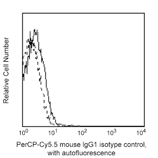
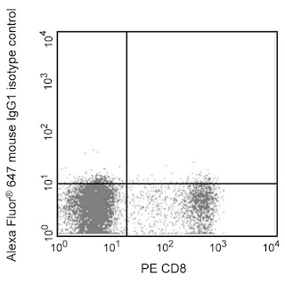
- An isotype control should be used at the same concentration as the antibody of interest.
- Caution: Sodium azide yields highly toxic hydrazoic acid under acidic conditions. Dilute azide compounds in running water before discarding to avoid accumulation of potentially explosive deposits in plumbing.
- Please observe the following precautions: Absorption of visible light can significantly alter the energy transfer occurring in any tandem fluorochrome conjugate; therefore, we recommend that special precautions be taken (such as wrapping vials, tubes, or racks in aluminum foil) to prevent exposure of conjugated reagents, including cells stained with those reagents, to room illumination.
- Alexa Fluor® 647 fluorochrome emission is collected at the same instrument settings as for allophycocyanin (APC).
- Alexa Fluor® is a registered trademark of Molecular Probes, Inc., Eugene, OR.
- The Alexa Fluor®, Pacific Blue™, and Cascade Blue® dye antibody conjugates in this product are sold under license from Molecular Probes, Inc. for research use only, excluding use in combination with microarrays, or as analyte specific reagents. The Alexa Fluor® dyes (except for Alexa Fluor® 430), Pacific Blue™ dye, and Cascade Blue® dye are covered by pending and issued patents.
- PerCP-Cy5.5–labelled antibodies can be used with FITC- and R-PE–labelled reagents in single-laser flow cytometers with no significant spectral overlap of PerCP-Cy5.5, FITC, and R-PE fluorescence.
- PerCP-Cy5.5 is optimized for use with a single argon ion laser emitting 488-nm light. Because of the broad absorption spectrum of the tandem fluorochrome, extra care must be taken when using dual-laser cytometers, which may directly excite both PerCP and Cy5.5™. We recommend the use of cross-beam compensation during data acquisition or software compensation during data analysis.
- For fluorochrome spectra and suitable instrument settings, please refer to our Multicolor Flow Cytometry web page at www.bdbiosciences.com/colors.
- Cy is a trademark of GE Healthcare.
- Please refer to www.bdbiosciences.com/us/s/resources for technical protocols.
Companion Products


Development References (6)
-
Annunziato F, Cosmi L, Liotta F, et al. Human Th17 cells: are they different from murine Th17 cells?. Eur J Immunol. 2009; 39:637-640. (Biology). View Reference
-
Annunziato F, Cosmi L, Santarlasci V, et al. Phenotypic and functional features of human Th17 cells. J Exp Med. 2007; 204:1849-1861. (Biology). View Reference
-
Sakaguchi, S, Yamaguchi, et al. Regulatory T cells and immune tolerance. Cell. 2008; 133(5):775-787. (Biology). View Reference
-
Shevach EM. Mechanisms of Foxp3+ T regulatory cell-mediated suppression. Immunity. 2009; 30:636-645. (Biology). View Reference
-
Zhu J, Paul WE. CD4 T cells: fates, functions, and faults. Blood. 2008; 112(5):1557-1569. (Biology). View Reference
-
Ziegler, SF. FOXP3: of mice and men. Annu Rev Immunol. 2006; 209-226. (Biology). View Reference
Please refer to Support Documents for Quality Certificates
Global - Refer to manufacturer's instructions for use and related User Manuals and Technical data sheets before using this products as described
Comparisons, where applicable, are made against older BD Technology, manual methods or are general performance claims. Comparisons are not made against non-BD technologies, unless otherwise noted.
For Research Use Only. Not for use in diagnostic or therapeutic procedures.