
Overview
While the COVID-19 global pandemic continues to evolve, BD Biosciences stands strong to support critical scientific research to better understand and ultimately battle the COVID-19 outbreak. Now more than ever, BD Biosciences is committed to being your scientific partner of choice and providing you with the tools and support required to enable your COVID-19 research. We provide a comprehensive portfolio of research tools to facilitate discovery in the following areas of COVID-19 research:
Viral immune response
Cytokine analysis
Vaccine research
Biomarkers and therapeutics
Navigate this site to learn more about BD Biosciences’ solutions and their applications in COVID-19 research. All products mentioned on these pages are for research use only, except for the BD FACSLyric™ Flow Cytometer, which is a combined-function flow cytometer for both IVD and research use. Its use in COVID-19 is for research purposes only.

For information on the comprehensive COVID-19 response efforts from BD—including product availability, status of BD operations and business continuity planning—visit bd.com/COVID-19.
Biology of SARS-CoV-2
COVID-19, or coronavirus disease 2019, is caused by the SARS-CoV-2 virus (severe acute respiratory syndrome coronavirus 2). SARS-CoV-2 is a novel virus belonging to the coronavirus family, which includes strains responsible for the common cold as well as the viruses responsible for SARS and MERS. It is genetically related to the coronavirus responsible for the SARS outbreak in 2003.
SARS-CoV-2 is an enveloped single-stranded RNA virus of the Coronaviridae family. Its genome encodes 29 proteins involved in the infection, replication and virion assembly process. Key structural proteins of the virus include the spike (S) glycoprotein, the envelope (E) protein and the matrix (M) transmembrane glycoprotein.1 Initial infection is believed to take place in nasal epithelial cells, which are rich in ACE-2 receptors.

Adapted from Callaway E, 2020.2
Viral entry also depends on cathepsin B/L activity and protease activity of TMPRSS2. These receptors are found distributed throughout epithelial cells of the respiratory and gastrointestinal tract. The S protein from SARS-CoV-2 enables cellular receptor binding, membrane fusion and hemagglutinin activity. The S protein contains a receptor binding domain (RBD), which is the most variable region of its genome. The RBD binds the human angiotensin-converting enzyme 2 (ACE-2) initiating viral uptake in human cells by endocytosis. The E and M proteins collaborate for viral envelope formation.1
Before entering a cell, SARS-CoV-2 needs to recognize and bind surface receptors such as ACE-2. From there, a cascade of sequential events take place to ensure successful viral infection and replication.2 The life cycle of SARS-CoV-2 includes the following:
SARS-CoV-2 binds to ACE-2 receptor
SARS-CoV-2 is endocytosed
SARS-CoV-2 fuses with a vesicle and viral RNA is released
SARS-CoV-2 RNA is translated into proteins
New SARS-CoV-2 virions are assembled
New SARS-CoV-2 virions are released
Types of immune responses to SARS-CoV-2
SARS-CoV-2 infection induces a multistep response from the host immune system:
First-line defense by the innate immune system with natural killer (NK) cells, macrophage activation and multinucleated giant cell formation
Proinflammatory cytokine and chemokine activation
Antigen presenting cells engulf and process SARS-CoV-2 antigens
T helper cells are activated by SARS-CoV-2 antigen
Cytotoxic T cells and B cells are cross activated by T helper cells
Immunological memory is formed for SARS-CoV-2
When the immune system fails to control its immune defense response properly, severe forms of COVID-19 develop.
Three major phases have been described in COVID-19 disease:
Viremia phase
Acute (pneumonia) phase
Recovery phase
Severe forms of SARS-CoV-2 induced COVID-19 are associated with increased release of pro-inflammatory cytokines (e.g., IL-6, Il-2, GM-CSF), also known as cytokine storm syndrome, hypercytokinemia or cytokine release syndrome, along with weak production of type I interferons (IFN-Is).3, 4
References
- Shang J, Wan Y, Luo C, et al. Cell entry mechanisms of SARS-CoV-2. Proc Natl Acad Sci U S A. 2020;117(21):11727-11734. doi:10.1073/pnas.2003138117
- Callaway E. The race for coronavirus vaccines. Nature. 2020;580(7805):576-577. doi: 10.1038/d41586-020-01221-y
- Sa Ribero M, Jouvenet N, Dreux M, Nisole S. Interplay between SARS-CoV-2 and the type I interferon response. PLoS Pathog. 2020;16(7):e1008737. doi:10.1371/journal.ppat.1008737
- Blanco-Melo D, Nilsson-Payant BE, Liu WC, et al. Imbalanced host response to SARS-CoV-2 drives development of COVID-19. Cell. 2020;181(5):1036-1045.e9. doi:10.1016/j.cell.2020.04.026
Scientific resources for COVID-19 research
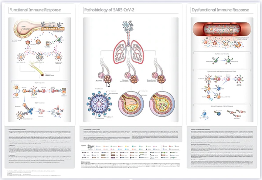
Immune Response to COVID-19
As the COVID-19 pandemic continues worldwide, there is a pressing need to understand the biological mechanisms determining COVID-19 disease progression and severity. Early research studies have reported dysfunctional immune responses and alterations in the immune cell composition in COVID-19 patients. The extent of the immune dysregulation appears to correlate with the severity of COVID-19 disease according to the literature. A deeper understanding of the interactions between SARS-CoV-2 and the host immune system will be important to further understand the pathogenesis of this disease and identify biomarkers that may potentially be used to predict and monitor COVID-19 progression.
The “COVID-19: Snapshot of the Immune Response” poster depicts the role different immune cells play in either a functional or dysfunctional immune response to SARS-CoV-2.
Multicolor Flow Cytometry Panels for COVID-19 Research
Flow cytometry can play a critical role in the study of COVID-19 immune response due to the ability to simultaneously measure expression of multiple proteins at the single-cell level. Flow cytometry is routinely used to assess immunophenotypic and functional changes (cytokine production, proliferation, cytotoxicity) in response to viral infection.
The “Multicolor Flow Cytometry Panels for COVID-19 Research” eBook and Interactive Cell Map tool contain a collection of multicolor flow cytometry panels and related protocols selected based on their relevance to the immunological changes observed in COVID-19 patients. The panels were designed using best panel-design practices and their performance, defined as the ability to clearly resolve populations of interest.
Monitoring the Immune System to Fight COVID-19
An overview of preliminary studies documenting the immunological changes observed in COVID-19 patients was recently presented by Dr. Maurice O’Gorman (Children's Hospital Los Angeles) and Dr. Andrea Cossarizza (University of Modena and Reggio Emilia School of Medicine).
Research Highlights
| Reference | Sample Size | Comparative Analysis | Immunological Changes |
|---|---|---|---|
Wang F, et al. The laboratory tests and host immunity of COVID-19 patients with different severity of illness. JCI Insight. 2020 May;5(10):e137799. doi: 10.1172/jci.insight.137799
| 65 COVID‑19 Patients | Extremely Severe vs Severe vs Moderate Cases | Total lymphocytes, total CD4+ and CD8+ T cells, B cells (absolute count) Total T cells, CD4+ cells (%); CD45RA+ Naïve Tregs (%) CD4+ CD28+ and CD8+ CD28+ T cells (%) CD86+ dendritic cells (%) CD4+ CD45RO+ memory T cells (%) HLA-DR+ activated CD4+ and CD8+ T cells (%) NK cells (%) PD-1+ exhausted CD4+ and CD8+ T cells (%) CD4+ and CD8+ T cells producing IFN-γ(%) Serum amounts of IL-6 and IL-10 (pg/mL) |
Chen G, et al. Clinical and immunological features of severe and moderate coronavirus disease 2019. J Clin Invest. 2020 May;130(5):2620-2629. doi: 10.1172/JCI137244 | 21 COVID‑19 Patients | Severe vs Moderate Cases | Total lymphocytes, CD4+ and CD8+ T cells (absolute count) Total B cells (%) CD45RA+ naïve Tregs (%) CD4+ T cells producing IFN-γ (%) Serum amounts of IL-6, IL-10, TNF, IL-2R (pg/mL) |
Qin C, et al. Dysregulation of immune response in patients with coronavirus 2019 (COVID-19) in Wuhan, China. Clin Infect Dis. 2020 Jul;71(15):762-768. doi: 10.1093/cid/ciaa248 | 452 COVID‑19 Patients | Severe vs Moderate Cases | Total lymphocytes, total T cells, CD4+ T cells (absolute count) CD4+ cells (%) CD4+ CD45RA+ naïve T cells (%) CD8+ CD28+ cells (%) Tregs (%) CD4+ CD45RO+ memory T cells (%) Serum amounts of TNF, IL-2R, IL-10, IL-6, IL-8 (pg/mL) |
Zheng M, et al. Functional exhaustion of antiviral lymphocytes in COVID-19 patients. Cell Mol Immunol. 2020 May;17(5):533-535. doi: 10.1038/s41423-020-0402-2 | 68 COVID‑19 Patients | Severe vs Moderate Cases | Total lymphocytes, CD8+ T cells and NK cells (absolute count) CD8+ T cells and NK cells expressing CD107a (%) CD8+ T cells and NK cells producing GZMB, IFN-γ, IL-2 and TNF (%) NKG2A+ exhausted CD8+ T cells and NK cells (%) |
Zheng H, et al. Elevated exhaustion levels and reduced functional diversity of T cells in peripheral blood may predict severe progression in COVID-19 patients. Cell Mol Immunol. 2020 May;17(5):541-543. doi: 10.1038/s41423-020-0401-3 | 6 Healthy Controls | Severe vs Moderate vs Healthy Cases | CD4+ T cells producing IFN-α or TNF (%) Polyfunctional CD8+ T cells producing IFN-γ, IL-2 and TNF (%) HLA-DR+ activated CD8+ T cells (%) PD-1+ CTLA-4+ TIGIT+ exhausted CD8+ T cells (%) |
Zhou Y, et al. Pathogenic T cells and inflammatory monocytes incite inflammatory storm in severe COVID-19 patients. Natl Sci Rev. 2020 June;7(6):998-1002. doi: 10.1093/nsr/nwaa041 | 33 COVID‑19 Patients | ICU vs No ICU Care | Total T cells, CD4+ and CD8+ T cells, monocytes, NK cells (absolute count) PD-1+TIM-3+ exhausted CD4+ and CD8+ T cells (%) CD4+ T cells producing IL-6 and GM-CSF (%) CD14+CD16+ monocytes producing IL-6 and GM-CSF (%) |
Diao B, et al. Reduction and functional exhaustion of T cells in patients with coronavirus disease 2019 (COVID-19). Front Immunol. May 2020. doi: 10.3389/fimmu.2020.00827 | 40 Healthy Subjects | ICU vs No ICU Care | Total T cells, CD4+ and CD8+ T cells (absolute count) PD-1+ exhausted CD4+ and CD8+ T cells (%) Serum amounts of IL-6, IL-10, TNF (pg/mL) |
Giamarellos-Bourboulis EJ, et al. Complex immune dysregulation in COVID-19 patients with severe respiratory failure. Cell Host Microbe. 2020 June;27(6):992-1000.e3. doi: 10.1016/j.chom.2020.04.009 | 54 COVID‑19 Patients | Macrophage Activated Syndrome vs Dysregulated Immune Response vs Intermediate Immune Response | Total CD4+ and CD8+ T cells, NK and B cells (absolute count) HLA-DR expression on monocytes (absolute number of molecules/cell) Neutrophils and macrophages (absolute count) Serum amounts of IL-6 (pg/mL) |
De Biasi S, et al. Marked T cell activation, senescence, exhaustion and skewing towards TH17 in patients with COVID-19 pneumonia. Nat Commun. 2020 July;11(1):3434. doi: 10.1038/s41467-020-17292-4 | 20 Healthy Subjects | COVID‑19 Patients vs Healthy Subjects | CD8+ naïve and central memory T cells (%) CD4+ and CD8+ TEMRA (%) HLA-DR+ CD38+ activated CD4+ and CD8+ T cells (%) PD-1+ CD57+ exhausted/senescent CD4+ and CD8+ T cells (%) Naïve Tregs (%) CD4+ and CD8+ T cells expressing CD107a (%) CD8+ T cells producing IL-2 or IL-17 (%) CD4+ T cells producing IL-2, IFN-α , TNF or IL-17 (%) Polyfunctional CD4+ T cells producing IL-2 and IL-17 (%) Serum amounts of CXCL6, IFN-γ, Galectin-1, -3, -9, IL-1α, IL-4, IL-6, IL-7, IL-8, IL-10, IL-15, IL-17, MICA, MIP-2 (CXCL2), CCL2, CCL3, CCL4, TNF (pg/mL) |
Maucourant C, et al. Natural killer cell immunotypes related to COVID-19 disease severity. Sci Immunol. 2020 August;5(50). doi: 10.1126/sciimmunol.abd6832 | 27 | Severe vs Moderate Cases | Total NK cells, CD56bright and CD56dim NK cells (absolute count) Ki67+ activated NK cells (%) CD57+NKG2C+ adaptive NK cells (%) |
Mathew D, et al. Deep immune profiling of COVID-19 patients reveals distinct immunotypes with therapeutic implications. Science. 2020 July;6508(369). doi: 10.1126/science.abc8511 | 60 Healthy Donors | COVID-19 Patients vs Recovered Donors vs Healthy Donors | T cells, CD4+ and CD8+ T cells (%) CD8+ effector memory RA T cells (%) CD4+ effector memory T cells (%) Ki67+ HLA-DR+ CD38+ activated CD4+ and CD8+ T cells (%) Activated circulating Tfh cells (%) CD27+CD38+ plasmablasts (%) Memory B cells (%) |
Sanchez-Cerillo I, et al. COVID-19 severity associates with pulmonary redistribution of CD1c+ DC and inflammatory transitional and nonclassical monocytes. J Clin Invest. 2020 August;140335. doi: 10.1172/JCI140335 | 22 Healthy Donors | Critical vs Severe vs Mild vs Healthy Cases | CD123+ plasmacytoid DCs (%) CD141+ conventional DCs (%) CD1c conventional DCs (%) Classical and nonclassical monocytes (%) |
Kuri-Cervantes L, et al. Comprehensive mapping of immune perturbations associated with severe COVID-19. Sci Immunol. 2020 Jul;5(49):eabd7114. doi: 10.1126/sciimmunol.abd7114 | 12 Healthy Donors | Severe vs Moderate vs Recovered vs Healthy Cases | CD4+ and CD8+ T cells, dendritic cells, CD56bright and CD56dim NK cells, MAIT cells and innate lymphoid cells (%) CD21+ CD27+ B cells (%) Neutrophiles, granulocytes and monocytes (%) HLA-DR+ CD38+ activated CD4+ and CD8+ T cells (%) CD27+CD38+ plasmablasts (%) Perforin+ granzyme+ cytotoxic CD8+ T cells (%) PD-1+ CD4+ T cells (%) CD21- CD27- B cells (%) |
The information on COVID-19 presented in the table above was compiled in October 2020.
The studies reported here have not been independently validated or verified by BD.
All BD reagents used in these studies are for Research Use Only. Not for use in diagnostic or therapeutic procedures.
Application data
BD Biosciences enables COVID-19 research by providing solutions for a comprehensive immunophenotypic, functional and transcriptional analysis of immune cells.
Flow Cytometry
- Assessment of immunophenotypic and functional changes in cells of the innate (e.g., monocytes, NK and dendritic cells) and adaptive (e.g., T and B cells) immune system
- Measurement of cytokines responsible for or associated with cytokine storm syndrome and/or hypercytokinemia
- Functional assays to assess antigen-specific T cell responses to viral epitopes
Fluorescence-Activated Cell Sorting
- Purification of antigen-specific T or B cells for vaccine efficacy testing or generation of neutralizing monoclonal antibodies
- Purification of immune cells of interest for downstream single-cell multiomic analysis
Single-Cell Multiomics
- Profiling immune cells of interest via targeted or whole transcriptome analysis
- Analysis of T or B cell receptor repertoire and assessment of cell clonality
- Enhanced immune cell characterization and signature discovery through simultaneous analysis of protein and mRNA
View product information, application data and scientific resources for assays performed on healthy human or murine cells that can also be potentially applicable to COVID-19 research.
Technology | Application | Description | Resources |
Flow cytometry | Immunophenotyping | Dried antibody cocktail for immunophenotypic characterization of human naïve, memory and effector T cells | |
Dried antibody cocktail for immunophenotypic characterization of human regulatory T cells | |||
Dried antibody cocktail for immunophenotypic characterization of human monocytes | |||
Assessment of markers associated with T cell exhaustion | |||
Immunophenotypic characterization of CD4+ T helper cells, Tregs and T follicular helper cells | |||
Immunophenotypic and functional characterization of polyfunctional T cells | |||
Immunophenotypic characterization of human NK cells | |||
Immunophenotypic characterization of human B cells | |||
Immunophenotypic characterization of murine dendritic cells | |||
Broad immunophenotypic characterization of major human immune cell lineages | |||
Broad immunophenotypic characterization of major murine immune cell lineages | |||
Functional assays | Proliferation, degranulation and intracellular cytokine analysis to assess CD8+ T cell activation | ||
Apoptosis, degranulation and intracellular cytokine analysis to assess NK cell cytotoxicity | |||
Intracellular cytokine analysis to assess the response of human dendritic cells to Toll-like Receptor agonists | |||
Quantitation of Th1/Th2/Th17 cytokines produced by activated T cells using BD® Cytometric Bead Array (CBA) | |||
Cell sorting and single-cell multiomics | Deep cell characterization and cell signature discovery | Identification of immunophenotypic and molecular signatures of chronically stimulated human T cells | |
Workflow for discovery and confirmation of cell signatures defining human innate lymphoid cell subsets | |||
Deep characterization of human regulatory T cells |
Reagent solutions for COVID-19 research
During this challenging time, researchers at the forefront of COVID-19 research need tools to power deeper cellular analysis. BD Biosciences research reagents include innovative dyes and a broad portfolio of human-specific antibodies to deliver more resolution and flexibility for experimental design, saving valuable samples and accelerating the time to critical insights.
Optimized Panels, Kits and Assays
Readily available optimized antibody cocktails, gene panels, kits and recommended multicolor flow cytometry panels shown in the tables below can help expedite experiments relevant to COVID-19 research.
| Research Area | Research Application | Product Solution(s) |
Viral Immune Response
| Identification of naïve and different memory T cell subsets in human whole blood | |
Identification of naïve and different effector regulatory T cell (Treg) subsets in human whole blood | ||
Identification of classical (M1), intermediate (M2) and nonclassical (M3) monocyte subsets in human whole blood | ||
Targeted human immune gene panel (399 genes) relevant to phenotyping human immune cells | ||
Targeted mouse immune gene panel (397 genes) relevant to phenotyping mouse immune cells | ||
Targeted human T cell gene panel (259 genes) relevant to T cell biology | ||
Whole transcriptome analysis (WTA) enables identification of the most differentially expressed genes in your samples. The WTA data can be used for cell type identification and to construct a targeted panel tailored for your samples of interest | ||
Cytokine Analysis
| Cytometric bead array to measure human cytokine(s) in serum, plasma and cell culture supernatant samples | BD® CBA Human Inflammatory Cytokine Kit BD® CBA Human Th1/Th2 Cytokine Kit |
Vaccine Research
| Detection of T and B cell clonotypes in samples of interest |
*Please contact your sales representative to request a quote.
| Research Area | Research Application | Recommended Research Panel | |||
| Viral Immune Response | Assess the expression of multiple T cell inhibitory receptors | T Cell Exhaustion Panel | |||
| Marker | Dye | Clone | Cat. No. | ||
CD3 | PerCP-Cy5.5 | UCHT1 | |||
CD4 | APC-R700 | RPA-T4 | |||
CD8 | APC-H7 | SK1 | |||
CD45RA | BV650 | HI100 | |||
CD197 (CCR7) | FITC | 150503 | |||
CD95 | BV480 | DX2 | |||
TIGIT | BV421 | 741182 | |||
CD152 (CTLA-4) | PE | BNI3 | |||
CD223 (LAG-3) | Alexa Fluor™ 647 | T47-530 | |||
CD272 (BTLA) | BV605 | J168-540 | |||
CD279 (PD-1) | PE-Cy7 | EH12.1 | |||
CD366 (TIM-3) | BV786 | 7D3 | |||
Identification of T helper cell (Th) subsets (Th1, Th2, Th17, Th22), follicular T helper cell (Tfh) subsets, regulatory T cell (Treg) subsets, and follicular regulatory T cell (Tfr) subsets | T Cell Subset Panel | ||||
| Marker | Dye | Clone | Cat. No | ||
Live/Dead | 7-AAD | N/A | |||
CD3 | Alexa Fluor™ 700 | UCHT1 | |||
CD4 | APC-H7 | SK3 | |||
CD45RA | BV605 | HI100 | |||
CD127 | BV711 | HIL-7R-M21 | |||
CD25 | BB515 | 2A3 | |||
CD183 (CXCR3) | PE-Cy7 | 1C6/CXCR3 | |||
CD185 (CXCR5) | BV480 | RF8B2 | |||
CD194 (CCR4) | BV421 | 1G1 | |||
CD196 (CCR6) | BV786 | 11A9 | |||
CD294 (CRTH2) | PE | BM16 | |||
CCR10 | APC | 1B5 | |||
BD Biosciences also offers a large and diverse portfolio of high-quality reagents to meet your specific experimental needs and enables the optimization and design of your own assay. Explore the availability of additional reagents relevant to different areas of COVID-19 research.
Immunophenotyping and Cell Sorting
The immune response to a virus such as SARS-CoV-2 can be tracked through cells and markers in the blood. Multicolor flow cytometry is one of the most powerful tools with which to identify, for example, alterations in the immune cell landscape in response to viral infection. BD Biosciences has a broad catalog of flow cytometry reagents to aid in the analysis and isolation of cells of the innate and adaptive immune system.
For example, T cells are a key component of viral immunity, not only indirectly fighting viral infections, but also in helping to mount a successful antibody response to provide lasting immunity. The table below shows a representative list of high-quality, flow cytometry–tested antibodies available in a broad range of colors that enable a comprehensive study of murine and human T cell biology.
| Type of Cell | CD8+ Cytotoxic T Lymphocytes (CTLs) | CD8+ Exhausted CTLs | CD4+ Th1 | CD4+ Th17 | CD4+ Tfh | CD4+ Tregs |
Main Function | Kill virus-infected cells | Activated but have lost cytotoxic activity | Promote cell-mediated response | Enhance neutrophil and macrophage recruitment | Regulate development of antigen specific B cell development and antibody production | Immune regulation |
Pathogens Targeted | Viruses and some intracellular pathogens | Intracellular pathogens | Extracellular pathogens | |||
Extracellular Markers | CD8 | CD279 (PD-1) | ||||
Differentiation Cytokines | ||||||
Hallmark Cytokines | ||||||
Transcription Factors |
Cytokine Response
Proper cytokine response is important for the clearance of any virus. In the case of COVID-19, early research suggests that overactive responses leading to cytokine release syndrome may contribute to more severe presentation of the disease.1,2 Some cytokines that have been identified as abnormally high in COVID-19 samples include:
BD Biosciences offers different solutions for comprehensive measurement of cytokine production, either at the single-cell (intracellular flow cytometry, BD FastImmune™ Cytokine Detection System) or bulk level (BD® Cytometric Bead Array, ELISA) for COVID-19 research.
BD FastImmune™ Cytokine Detection System
With streamlined and highly reproducible intracellular cytokine staining, the BD FastImmune™ System is designed to meet the needs of applied research on human samples. As a complete and optimized system, it is particularly well suited for research studies assessing immune responses to virus or vaccine candidates.
BD Biosciences provides a range of BD FastImmune™ Multicolor or Single-Color Cytokine Reagents, produced under RUO (GMP) conditions, for antibody staining, including selected sample-activation and sample-processing reagents.
Available cocktails:
Selected list of relevant single-color BD FastImmune™ Reagents for analysis of cytokine and degranulation marker expression.
BD® Cytometric Bead Array (CBA)
BD® CBA Solutions are available in preconfigured kits or in an open, configurable format for unique needs.
BD® CBA Flex Sets provide an open and configurable method of detection so that researchers can build their own multiplexes for protein quantitation as low as 10 pg/mL.
BD® CBA Enhanced Sensitivity Flex Sets are capable of detecting cytokine concentrations as low as 0.274 pg/mL.
BD® CBA Kits are preconfigured for achieving consistent results for routine panels.
Selected list of relevant BD® CBA Flex Sets:
ELISAs
BD Biosciences offers three convenient ELISA reagent formats for quantitation of cytokines and other soluble proteins.
Individual reagents are composed of unlabeled capture antibodies, biotinylated detection antibodies and protein standards offering a wide range of specificities and assay design flexibility. BD OptEIA™ Sets include reagents for five or twenty 96-well plates and pre-coated 96-well plates. All three BD Biosciences sandwich ELISA formats provide fast, easy quantitation of soluble analytes and total response analysis.
Selected list of relevant BD OptEIA™ ELISA Sets:
BD® AbSeq Antibodies
Utilize the cutting-edge antibody-oligo technology in the form of BD® AbSeq Antibodies to probe up to 100 different cell surface proteins at a single-cell level, while simultaneously performing single-cell RNA analyses. Multiomic research using BD® AbSeq Antibodies enables deeper characterization of your immune populations of interest and the potential discovery of novel cell immune signatures.
References
- Shi Y, Wang Y, Shao C, et al. COVID-19 infection: the perspectives on immune responses. Cell Death Differ. 2020;27(5):1451-1454. doi: 10.1038/s41418-020-0530-3.
- Cossarizza A, De Biasi S, Guaraldi G, Girardis M, Mussini C. SARS-CoV-2, the virus that causes COVID-19: cytometry and the new challenge for global health. Cytometry A. 2020;97(4):340-343. doi: 10.1002/cyto.a.24002.
Instrument solutions for COVID-19 research
Cell Analyzers
Flow cytometry has been a trusted approach to analyze cells and sort them to near-purity (~98%) for decades, and more recently, the analysis of small entities such as virus, exosomes and extracellular vesicles have become more mainstream. BD Biosciences portfolio of cell analyzers offers the solutions necessary for your COVID-19 related research experiments.
BD FACSymphony™
Offering customized solutions for high-parameter analysis, the BD FACSymphony™ Cell Analyzers can scale up to meet your multicolor needs. When your SARS-CoV-2 research relies on either broad or deep high-parameter phenotypic characterization of specific subpopulations of immune cells, rely on the BD FACSymphony™ Analyzer.

BD FACSLyric™
The BD FACSLyric™ Flow Cytometer is upgradeable from 4 to 12 colors and offers high resolution and simplified standardization across instruments and labs, enabling collaboration and accelerating translational research. These make the BD FACSLyric™ Flow Cytometer a powerful tool for COVID-19 research. For more information on the research use of the BD FACSLyric™ Flow Cytometer contact your local representative.

BD FACSCelesta™
The BD FACSCelesta™ Flow Cytometer is designed to make high-performance, multicolor flow cytometry accessible to individual labs analyzing up to 14 parameters. Drive your fundamental research in viral immune response forward without waiting for the core lab.

Cell Sorters
BD has led the way for 40 years in cell sorting. BD sorters combined with custom biosafety cabinets with integrated aerosol management systems from The Baker Company have been verified to provide product and personnel protection according to several bioprotection standards. In addition, the BD FACSAria™ product family and BD FACSymphony™ S6 Containment Systems have been verified using the recommended Cyclex-d/Dragon Green microsphere assay.1 Information about a new SARS-CoV-2 sorting protocol is available at the ISAC Biosafety website.
BD FACSymphony™ S6
Building on the BD FACSAria™ Fusion biosafety solution, the BD FACSymphony™ S6 Cell Sorter adds high-parameter analysis to isolate populations based on more than 20 parameters and 6-way sorting capabilities to isolate more populations from one precious sample. Transfer panels from the BD FACSymphony™ A5 Cell Analyzer to isolate ultraspecific immune cell subpopulations with complex phenotypes in order to research antigen-specific cell subsets.
Additional resources:
Microbiological Test Report – BD FACSymphony™ S6 Cell Sorter

BD FACSMelody™
A truly easy-to-use cell sorter for your own lab, the BD FACSMelody™ with 4-way sorting capability makes the complex world of flow cytometry and sorting available to more researchers. It combines proven BD technology with new automation and simplified software to deliver high-performance results in less time. The BD FACSMelody™ Cell Sorter provides an easy solution for purification of immune cells of interest for downstream functional or multiomic assays for research use.
Additional resources:
Microbiological Test Report – BD FACSMelody™ Cell Sorter

BD FACSAria™ Fusion
The BD FACSAria™ Fusion Cell Sorter delivers exceptional multicolor performance and ease-of-use, combined with best-in-class biosafety expertise for an advanced cell sorter and biosafety solution. Purification of antigen-specific T and B cells for downstream functional assays is a major application in vaccine research, and the BD FACSAria™ Fusion Cell Sorter is built to deliver on this front.
Additional resources:
Microbiological Test Report – BD FACSAria™ Fusion Cell Sorter

Single-Cell Multiomics
BD Rhapsody™ Single-Cell Analysis System
Perform multiomic analyses by simultaneously probing for protein and RNA expression in immune cells with the BD Rhapsody™ Single-Cell Analysis System. The multiomic data generated may unlock discoveries of novel immune subsets—enabling the identification molecular drivers of immune cell activation and exhaustion in response to viral infections. Identification and tracking of clonotypes in response to a vaccine may also be achieved through T and B cell receptor repertoire analysis leveraging the BD single-cell solution.

Reference
Perfetto SP, Hogarth PJ, Monard S, et al. Novel impactor and microsphere-based assay used to measure containment of aerosols generated in a flow cytometer cell sorter. Cytometry A. 2019;95(2):173-182. doi: 10.1002/cyto.a.23680.
Data analysis
BD Biosciences offers powerful informatics tools to complement your flow cytometry and multiomic analysis. These workflows are used by scientists in the lab to enable data insights in infectious disease and vaccine research. Supporting scientific research is our top priority.
Flow Cytometry Analysis
FlowJo™ Software is the leading platform for single-cell flow cytometry analysis that helps your research stand out with accessible features for immunophenotyping, cell cycle, proliferation, kinetics studies, quantitative population comparison, plate screening assays and more.

RNA-Seq Analysis
SeqGeq™ v1.6 Software analyzes data from RNA-Seq and single-cell RNA-Seq experiments, providing tools to extract genes and cells of interest and putting transcriptome analyses back in the hands of bench scientists. Explore, visualize and shape your next-generation sequencing data with interactive graphing tools that are designed to help you focus on the insights that matter most.

We offer a variety of support and training for when you need it most:
BD Cares
As a result of the COVID-19 pandemic, the world of healthcare as we know it has changed, and hospitals and laboratories are facing unprecedented challenges as a result.
At BD, we take our role as your science partner of choice seriously, and we understand that whether you’re on the frontlines, working in a laboratory behind the scenes, or doing your part by socially distancing, the role you play is invaluable. Now more than ever, scientific discoveries and clinical research must continue to flow.
We are here to support you every step of the way:
- Enhancing instrument support capabilities. For a limited time, we are offering complimentary remote instrument diagnostics and serviceability through BD Assurity Linc™ Remote Systems Management Software in addition to virtual instrument training.
- Increasing opportunities to further your flow cytometry knowledge and earn CE credit through virtual educational webinars.
- Providing limited-time special offers on instrument and reagents.
Together, we will make it through this pandemic and emerge strong.
BD Assurity Linc™ Remote Systems Management Software
In times of limited onsite interactions due to COVID-19 restrictions, BD has reinforced its remote support capabilities. BD Assurity Linc™ Remote Services, now powered by BD RSS technology, provides remote access into BD systems, allowing BD Service and Support teams to monitor your instruments and address technical issues remotely in a timely manner.
Enjoy the benefits of reduced service visits, faster issue resolution and high-level security.
If you are interested in installing this software or learning more, please contact GMB-AssurityLinc@bd.com.
Remote instrument diagnostics and serviceability through BD Assurity Linc™ Remote Services
BD Assurity Linc™ Remote Services enables the BD team to remotely monitor your instruments, address technical issues and potentially repair your instruments—with no on-site service visit required. In an effort to keep our associates, customers and communities safe, BD is offering this service at no cost to you for a limited time.
Contact GMB-AssurityLinc@bd.com to activate your instrument.
Virtual instrument training and troubleshooting
A dedicated team of 200+ BD associates is available to remotely address your needs. We have the required tools to execute virtual instrument training and support—whether it’s through FaceTime, Microsoft Teams or other channels.
On-site support
Where our on-site support is needed to ensure immediate patient care, such as technology installation/service and clinical support, BD Field Service Engineers will be there at your request. We are taking precautions to protect our associates, customers and patients by following guidance from the World Health Organization, U.S. Centers for Disease Control and each of our customers’ guidelines on access to their facilities.
Education
Further your flow cytometry knowledge through the BD library of on-demand webinars and upcoming virtual events.
Limited-Time Special Offers
To help navigate potential financial constraints placed upon your institutions, BD is providing various limited-time special offers for US customers. Visit the BD promotions webpage or contact your local sales representative for the latest offers.
Class 1 Laser Product.
For Research Use Only. Not for use in diagnostic or therapeutic procedures.
Alexa Fluor is a trademark of Life Technologies Corporation.
Cy is a trademark of Global Life Sciences Solutions Germany GmbH or an affiliate doing business as Cytiva.
FaceTime is a trademark of Apple Inc. Microsoft Teams is a trademark of Microsoft Corporation.