-
Training
- Flow Cytometry Basic Training
-
Product-Based Training
- FACSAria Product Based Training
- FACSMelody Product-Based Training
- FACSLyric Product-Based Training
- FACSCanto Product-Based Training
- LSRFortessa Product-Based Training
- FACSymphony Product-Based Training
- FACSDuet Product-Based Training
- HTS Product-Based Training
- BD FACSDiscover™ S8 Cell Sorter Product Training
-
Advanced Training
-
Thought Leadership
-
Product News
- Blogs
- Scientific Publications
-
Events
- Nature Research Academies Workshop 2023
- CYTO 2023: Advancing the World of Cytometry
- Singapore Gene & Cell Therapy Conference 2023
- EuroFlow Educational Workshop
- Nature Research Masterclass 2023
- Novel Approaches to Single-Cell Plant Research: from Real-Time Imaging Cell Sorting to Single-Nuclei Transcriptomics
- Advances in Immune Monitoring Series
-
Product News
-
- FACSAria Product Based Training
- FACSMelody Product-Based Training
- FACSLyric Product-Based Training
- FACSCanto Product-Based Training
- LSRFortessa Product-Based Training
- FACSymphony Product-Based Training
- FACSDuet Product-Based Training
- HTS Product-Based Training
- BD FACSDiscover™ S8 Cell Sorter Product Training
-
- Nature Research Academies Workshop 2023
- CYTO 2023: Advancing the World of Cytometry
- Singapore Gene & Cell Therapy Conference 2023
- EuroFlow Educational Workshop
- Nature Research Masterclass 2023
- Novel Approaches to Single-Cell Plant Research: from Real-Time Imaging Cell Sorting to Single-Nuclei Transcriptomics
- Advances in Immune Monitoring Series
- Singapore (English)
-
Change country/language
Old Browser
This page has been recently translated and is available in French now.
Looks like you're visiting us from {countryName}.
Would you like to stay on the current country site or be switched to your country?
BD Horizon™ CFSE
(RUO)


Flow cytometric analysis of proliferating peripheral blood lymphocytes stained with BD Horizon™ CFSE. Human PBMC were labeled (10 min, 37°C) with BD Horizon™ CFSE (Cat. No. 565082; 2.5 μM). Cells were washed twice and cultured (4 days) alone (dashed line histogram) or with (solid line histogram) phytohemagglutinin. The cells were stained with 0.1 μg/mL DAPI (Cat. No. 564907) to exclude nonviable cells. The histogram shows CFSE fluorescence peaks of gated events with the light-scatter characteristics of viable lymphocytes (DAPI-negative). Unstimulated cells show a single, bright CFSE fluorescence peak, indicating no proliferation. Stimulated cells show multiple CFSE fluorescence peaks, indicating multiple generations of proliferating cells. Analysis was performed using a BD LSRFortessa™ Cell Analyzer System.

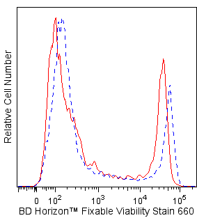
Two-color flow cytometric analysis of proliferating mouse splenocytes stained with BD Horizon™ CFSE. BALB/c mouse spleen cells were treated with BD Pharm Lyse™ Lysing Buffer (Cat. No. 555899) to lyse erythrocytes. The splenic leucocytes were washed and labeled with BD Horizon™ CFSE (1 μM) for 10 minutes at 37°C. The cells were washed twice and then cultured with Purified NA/LE Hamster Anti-Mouse CD3e (Cat. No. 553057) and Purified NA/LE Hamster Anti-Mouse CD28 (Cat. No. 553294) antibodies. After 3 days, the cells were harvested, washed, and restimulated (4 hrs) with Phorbol 12-Myristate 13-Acetate and Ionomycin in the presence of BD GolgiStop™ Protein Transport Inhibitor (containing Monensin) (Cat. No. 554724). Cells were harvested, stained with BD Horizon™ Fixable Viability Stain 660 (Cat. No. 564405; to exclude nonviable cells), fixed, permeabilized using a BD Cytofix/Cytoperm™ Fixation/Permeabilization Solution Kit (Cat. No. 554714), and then stained with PE Anti-Mouse CD4 (Cat. No. 553048/553049/561837) and PE-Cy™7 Anti-Mouse IL-2 (Cat. No. 560538) antibodies. Two-color flow cytometric dot plots showing the correlated expression patterns of CFSE versus CD4 (Left Panel) or IL-2 (Right Panel) were derived from gated events with the forward and side light-scatter characteristics of viable lymphocytes. Flow cytometric analysis was performed using a BD LSRFortessa™ Cell Analyzer System.

BD Horizon™ CFSE

BD Horizon™ CFSE
Regulatory Status Legend
Any use of products other than the permitted use without the express written authorization of Becton, Dickinson and Company is strictly prohibited.
Preparation And Storage
Recommended Assay Procedures
Materials Provided
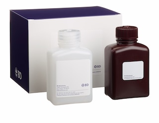
CFSE dye (2 vials, 500 μg dye per vial)
Materials needed but not provided
• Viable single-cell suspensions of primary lymphoid cells of interest
• Fresh cell culture-grade Dimethyl Sulfoxide (DMSO; eg, Sigma D2650)
• Sterile Dulbecco's Phosphate Buffered Saline (1× DPBS)
• Cell culture medium, eg, with 10% Fetal Bovine Serum (FBS)
• Sterile polypropylene 15-mL or 50-mL conical tubes
• Microcentrifuge tubes or 2-mL cryovials
• Fluorescent antibodies for immunophenotypic or functional analysis (optional)
Required Equipment
• Blue laser-equipped cytometer; eg, BD FACSCanto™ II, BD LSRFortessa™, BD LSR™ II Flow Cytometer, or BD Accuri™ C6
• Centrifuge
• 37°C CO2 Incubator and 37ºC Water Bath
Storage
Upon arrival, store the dye powder with desiccant and protected from light at ≤ -20°C until use. After reconstitution with DMSO, store the stock solution with desiccant at ≤ -20°C in small aliquots. The stock solution is stable for 3 months if stored as indicated.
Procedure
CFSE has a maximum absorption of 490 nm and a maximum emission of 520 nm. Before staining with this reagent, please confirm that your flow cytometer is capable of exciting the fluorochrome and discriminating the emitted fluorescence.
Preparation of CFSE dye solution in DMSO
1. Prepare a 10 mM stock solution of CFSE dye by adding 90 μL DMSO to one vial of CFSE. Mix thoroughly by vortexing.
2. Prepare single-use aliquots of the CFSE stock solution (user-defined volume) using appropriately labeled microcentrifuge tubes or 2-mL cryovials.
3. Freeze all aliquots at ≤ -20°C with desiccant and protected from light.
CFSE labeling of cells
This procedure has been optimized for labeling mouse and human lymphocytes at cell concentrations of 10-30 × 10^6 cells/mL. Assay conditions may need to be optimized for other cell types or cell densities.
1. Thaw the 10 mM stock solution of CFSE, if previously frozen.
2. Transfer cells to 15- or 50-mL polypropylene centrifuge tubes.
3. Wash cells in 1× DPBS to remove any residual serum proteins.
4. Repeat step 3.
5. Resuspend the cells thoroughly into a single cell suspension at a concentration of 10-30 × 10^6 cells/mL in 1× DPBS.
a. Note: Avoid staining in azide-, serum-, or protein-containing buffers, as these moieties bind free dye and may interfere with staining of the cells.
6. Add stock CFSE to the single cell suspension to achieve a final concentration of 1-10 μM. If necessary, the stock solution may be diluted further with DMSO before addition to the single cell suspension. Vortex immediately.
7. Incubate the dye-cell suspension in a 37°C water bath for 10-15 minutes.
8. Add 9× the original volume of 1× DPBS to the cells and pellet cells by centrifugation.
9. Decant the supernatant and gently mix to disrupt the cell pellet.
10. Add 10 mL of complete medium with 10% FBS and repeat centrifugation step.
11. Decant supernatant and gently mix to disrupt the cell pellet.
12. Resuspend the cells in complete medium and proceed to cell culture or analysis by flow cytometry.
Notes:
1. Confirm brightness of the cell labeling by comparing labeled cells with unlabeled control cells.
2. BD Horizon™ CFSE can be used in intracellular staining assays that require fixation with formaldehyde and permeabilization with methanol and detergents such as those used for BD Phosflow™ staining (eg, Cat. No. 558050, BD Phosflow™ Perm Buffer III), intracellular cytokine staining (eg, Cat. No. 554714, BD Cytofix/Cytoperm™ Fixation/Permeablization Kit), or transcription factor staining (eg, Cat. No. 562574/562725, BD Pharmingen™ Transcription Factor Buffer Set).
3. Higher initial staining intensities can be achieved by increasing the initial CFSE dye loading concentration. However, as with other succinimidyl ester containing tracking dyes, higher loading concentrations may lead to increased cell toxicity or cell death.
Product Notices
- Since applications vary, each investigator should titrate the reagent to obtain optimal results.
- For fluorochrome spectra and suitable instrument settings, please refer to our Multicolor Flow Cytometry web page at www.bdbiosciences.com/colors.
- Cy is a trademark of GE Healthcare.
- Before staining with this reagent, please confirm that your flow cytometer is capable of exciting the fluorochrome and discriminating the resulting fluorescence.
- Please refer to www.bdbiosciences.com/us/s/resources for technical protocols.
Companion Products


BD Horizon™ CFSE (carboxyfluorescein diacetate succinimidyl ester) is a blue laser excitable dye that can be used for flow cytometric monitoring of cell divisions. The non-fluorescent dye passively diffuses across cell membranes and is cleaved by intracellular esterases within viable cells. The cleaved dye becomes highly fluorescent and covalently binds to protein amine groups within the cells via its succinimidyl ester group. Nonviable cells remain nonfluorescent. As viable cells divide, CFSE dye is distributed uniformly between daughter cells; each daughter cell retains approximately half of the CFSE fluorescence intensity of its parent cell. The covalently bound dye is well retained within the cells allowing for applications involving live or aldehyde-fixed and permeabilized cells.
BD Horizon™ CFSE has an excitation maximum of 490 nm and an emission maximum of 520 nm.
Development References (4)
-
Lyons AB, Blake SJ, Doherty KV. Flow cytometric analysis of cell division by dilution of CFSE and related dyes. Curr Protoc Cytom. 9(9.11)(Methodology). View Reference
-
Lyons AB, Doherty KV. Flow cytometric analysis of cell division by dye dilution. Curr Protoc Cytom. 2004; 9(Unit 9.11):1-9. (Methodology). View Reference
-
Lyons AB, Parish CR. Determination of lymphocyte division by flow cytometry. J Immunol Methods. 1994; 171(1):131-137. (Methodology). View Reference
-
Lyons AB. Analysing cell division in vivo and in vitro using flow cytometric measurement of CFSE dye dilution. J Immunol Methods. 2000; 243(1-2):147-154. (Methodology). View Reference
Please refer to Support Documents for Quality Certificates
Global - Refer to manufacturer's instructions for use and related User Manuals and Technical data sheets before using this products as described
Comparisons, where applicable, are made against older BD Technology, manual methods or are general performance claims. Comparisons are not made against non-BD technologies, unless otherwise noted.
For Research Use Only. Not for use in diagnostic or therapeutic procedures.
Report a Site Issue
This form is intended to help us improve our website experience. For other support, please visit our Contact Us page.