Preparation And Storage
Product Notices
- This reagent has been pre-diluted for use at the recommended Volume per Test. We typically use 1 × 10^6 cells in a 100-µl experimental sample (a test).
- An isotype control should be used at the same concentration as the antibody of interest.
- Source of all serum proteins is from USDA inspected abattoirs located in the United States.
- Caution: Sodium azide yields highly toxic hydrazoic acid under acidic conditions. Dilute azide compounds in running water before discarding to avoid accumulation of potentially explosive deposits in plumbing.
- For fluorochrome spectra and suitable instrument settings, please refer to our Multicolor Flow Cytometry web page at www.bdbiosciences.com/colors.
- Please refer to http://regdocs.bd.com to access safety data sheets (SDS).
- Please refer to www.bdbiosciences.com/us/s/resources for technical protocols.
Companion Products


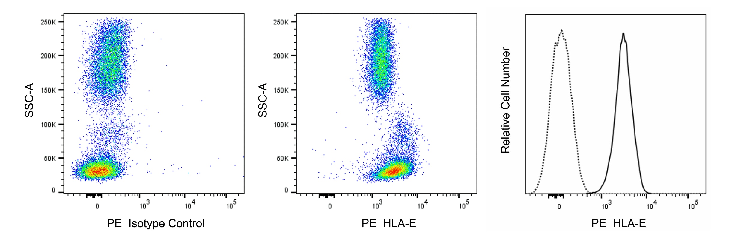
The 3D12 monoclonal antibody specifically recognizes Human Leukocyte Antigen E (HLA-E) that is widely expressed on leucocytes and some other cell types. Cell surface HLA-E is normally expressed as a noncovalent complex comprised of the ~45 kDa type I transmembrane, HLA-E heavy-chain glycoprotein, the ~12 kDa invariant β2-microglobulin (β2m) light chain, and a short bound peptide. Human HLA-E represents a nonclassical Major Histocompatibility Complex class I (MHC class Ib) molecule that is homologous to mouse H-2 Qa-1. Although structurally related to the classical, highly polymorphic MHC class Ia antigens (HLA-A, -B, -C), HLA-E shows limited polymorphism. HLA-E functions in the regulation or self-nonself discrimination of innate and adaptive immune responses. In addition to binding self peptides, the HLA-E complex can selectively bind and present peptides derived from bacterial or viral pathogen-infected cells, stressed cells, or tumor cells to elicit antigen-specific, HLA-E-restricted CD8+ T cell responses. The cell surface HLA-E complex likewise serves as a ligand for heterodimeric CD94:NKG2A inhibitory and CD94:NKG2C activating receptors that are differentially expressed on NK cells and some T cells. These ligand:receptor interactions can either suppress or promote NK or T cell-mediated responses. The 3D12 antibody reportedly binds to both free or complexed HLA-E heavy chain and can block HLA-E-dependent function.

Development References (6)
-
Joosten SA, Sullivan LC, Ottenhoff TH. Characteristics of HLA-E Restricted T-Cell Responses and Their Role in Infectious Diseases. J Immunol Res. 2016; 2016:1-11. (Biology). View Reference
-
Joosten SA, van Meijgaarden KE, van Weeren PC, et al. Mycobacterium tuberculosis peptides presented by HLA-E molecules are targets for human CD8 T-cells with cytotoxic as well as regulatory activity.. PLoS Pathog. 2010; 6(2):e1000782. (Clone-specific: Flow cytometry). View Reference
-
Lee N, Goodlett DR, Ishitani A, Marquardt H, Geraghty DE. HLA-E surface expression depends on binding of TAP-dependent peptides derived from certain HLA class I signal sequences.. J Immunol. 1998; 160(10):4951-60. (Immunogen: Flow cytometry, Immunoprecipitation, Western blot). View Reference
-
Lee N, Llano M, Carretero M, et al. HLA-E is a major ligand for the natural killer inhibitory receptor CD94/NKG2A.. Proc Natl Acad Sci USA. 1998; 95(9):5199-204. (Clone-specific: Functional assay, Immunoprecipitation, Western blot). View Reference
-
Llano M, Lee N, Navarro F, et al. HLA-E-bound peptides influence recognition by inhibitory and triggering CD94/NKG2 receptors: preferential response to an HLA-G-derived nonamer.. Eur J Immunol. 1998; 28(9):2854-63. (Clone-specific: Flow cytometry, Functional assay). View Reference
-
Ogg G, Cerundolo V, McMichael AJ. Capturing the antigen landscape: HLA-E, CD1 and MR1.. Curr Opin Immunol. 2019; 59:121-129. (Biology). View Reference
Please refer to Support Documents for Quality Certificates
Global - Refer to manufacturer's instructions for use and related User Manuals and Technical data sheets before using this products as described
Comparisons, where applicable, are made against older BD Technology, manual methods or are general performance claims. Comparisons are not made against non-BD technologies, unless otherwise noted.
For Research Use Only. Not for use in diagnostic or therapeutic procedures.