





.png)


Preparation And Storage


Recommended Assay Procedures
The FITC-conjugated MAb11 antibody can be used for multicolor immunofluorescent staining and flow cytometric analyses to identify and enumerate TNF-producing cells within mixed cell populations (see figure). For optimal immunofluorescent staining with flow cytometric analysis, this anti-cytokine antibody should be titrated (≤ 0.5 µg mAb/million cells) For specific methodology, please visit the protocols section under "Cytokines (Intracellular Staining)" or "Intracellular Flow" at our website, http://www.bdbiosciences.com/us/s/resources.
A useful control for demonstrating specificity of staining is either of the following: 1) pre-block the conjugated MAb11 antibody with a molar excess of ligand (e.g., recombinant human TNF; Cat No. 554618) prior to staining, or 2) pre-block the fixed/ permeabilized cells with unlabelled MAb11 antibody (Cat. No. 554510) prior to staining. The staining technique and blocking controls are described in detail by C. Prussin and D. Metcalfe. A suitable mouse IgG1 isotype control for assessing the level of background staining on paraformaldehyde-fixed/saponin-permeabilized human cells is FITC-MOPC-21 (Cat. No. 554679); use at comparable concentrations to antibody of interest.
Product Notices
- Since applications vary, each investigator should titrate the reagent to obtain optimal results.
- An isotype control should be used at the same concentration as the antibody of interest.
- Caution: Sodium azide yields highly toxic hydrazoic acid under acidic conditions. Dilute azide compounds in running water before discarding to avoid accumulation of potentially explosive deposits in plumbing.
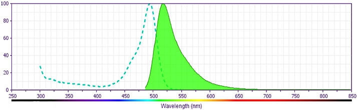
- For fluorochrome spectra and suitable instrument settings, please refer to our Multicolor Flow Cytometry web page at www.bdbiosciences.com/colors.
- Species cross-reactivity detected in product development may not have been confirmed on every format and/or application.
- Please refer to www.bdbiosciences.com/us/s/resources for technical protocols.
Companion Products


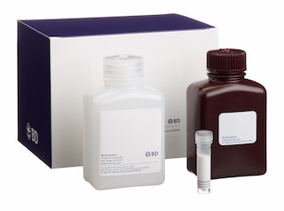
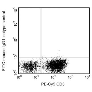
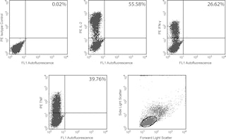
The MAb11 monoclonal antibody specifically binds to human tumor necrosis factor (TNF, also known as TNF-α) protein. TNF is an efficient juxtacrine, paracrine and endocrine mediator of inflammatory and immune functions. It regulates the growth and differentiation of a variety of cell types. TNF is cytotoxic for transformed cells when in conjunction with IFN-γ. It is secreted by activated monocytes/macrophages and other cells such as B cells, T cells and fibroblasts. The immunogen used to generate the MAb11 hybridoma was recombinant human TNF. The MAb11 antibody has been reported to crossreact with Rhesus Macaque TNF.
Development References (10)
-
Andersson J, Abrams J, Bjork L, et al. Concomitant in vivo production of 19 different cytokines in human tonsils. Immunology. 1994; 83(1):16-24. (Biology). View Reference
-
Andersson U, Andersson J. Immunolabeling of cytokine-producing cells in tissues and in suspension. In: Fradelizie D, Emelie D, ed. Cytokine Producing Cells. Paris: Inserm; 1994:32-49.
-
Danis VA, Franic GM, Rathjen DA, Brooks PM. Effects of granulocyte-macrophage colony-stimulating factor (GM-CSF), IL-2, interferon-gamma (IFN-gamma), tumour necrosis factor-alpha (TNF-alpha) and IL-6 on the production of immunoreactive IL-1 and TNF-alpha by human monocytes. Clin Exp Immunol. 1991; 85(1):143-150. (Clone-specific: ELISA). View Reference
-
Jason J, Larned J. Single-cell cytokine profiles in normal humans: comparison of flow cytometric reagents and stimulation protocols. J Immunol Methods. 1997; 207(1):13-22. (Biology). View Reference
-
Litton M, Andersson J, Bjork L, Fehniger T, Ulfgren AK, Andersson U. Cytoplasmic cytokine staining in individual cells. In: Debets and Savelkoul, ed. Human Cytokine Protocols. Humana Press; 1996.
-
Prussin C, Metcalfe DD. Detection of intracytoplasmic cytokine using flow cytometry and directly conjugated anti-cytokine antibodies. J Immunol Methods. 1995; 188(1):117-128. (Methodology). View Reference
-
Raqib R, Lindberg AA, Wretlind B, Bardhan PK, Andersson U, Andersson J. Persistence of local cytokine production in shigellosis in acute and convalescent stages. Infect Immun. 1995; 63(1):289-296. (Biology). View Reference
-
Rathjen DA, Cowan K, Furphy LJ, Aston R. Antigenic structure of human tumour necrosis factor: recognition of distinct regions of TNF alpha by different tumour cell receptors. Mol Immunol. 1991; 28(1-2):79-86. (Clone-specific: ELISA). View Reference
-
Sander B, Andersson J, Andersson U. Assessment of cytokines by immunofluorescence and the paraformaldehyde-saponin procedure. Immunol Rev. 1991; 119:65-93. (Biology). View Reference
-
Verdier F, Aujoulat M, Condevaux F, Descotes J. Determination of lymphocyte subsets and cytokine levels in cynomolgus monkeys. Toxicology. 1995; 105(1):81-90. (Biology). View Reference
Please refer to Support Documents for Quality Certificates
Global - Refer to manufacturer's instructions for use and related User Manuals and Technical data sheets before using this products as described
Comparisons, where applicable, are made against older BD Technology, manual methods or are general performance claims. Comparisons are not made against non-BD technologies, unless otherwise noted.
For Research Use Only. Not for use in diagnostic or therapeutic procedures.
