Old Browser
This page has been recently translated and is available in French now.
Looks like you're visiting us from {countryName}.
Would you like to stay on the current country site or be switched to your country?


Regulatory Status Legend
Any use of products other than the permitted use without the express written authorization of Becton, Dickinson and Company is strictly prohibited.
Preparation And Storage
Recommended Assay Procedures
BD™ CompBeads can be used as surrogates to assess fluorescence spillover (Compensation). When fluorochrome conjugated antibodies are bound to BD CompBeads, they have spectral properties very similar to cells. However, for some fluorochromes there can be small differences in spectral emissions compared to cells, resulting in spillover values that differ when compared to biological controls. It is strongly recommended that when using a reagent for the first time, users compare the spillover on cells and BD CompBead to ensure that BD CompBeads are appropriate for your specific cellular application.
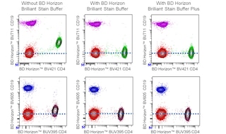
For optimal and reproducible results, BD Horizon Brilliant Stain Buffer should be used anytime two or more BD Horizon Brilliant dyes are used in the same experiment. Fluorescent dye interactions may cause staining artifacts which may affect data interpretation. The BD Horizon Brilliant Stain Buffer was designed to minimize these interactions. More information can be found in the Technical Data Sheet of the BD Horizon Brilliant Stain Buffer (Cat. No. 563794/566349) or the BD Horizon Brilliant Stain Buffer Plus (Cat. No. 566385).
Note: When using high concentrations of antibody, background binding of this dye to erythroid cell subsets (mature erythrocytes and precursors) has been observed. For researchers studying these cell populations, or in cases where light scatter gating does not adequately exclude these cells from the analysis, this background may be an important factor to consider when selecting reagents for panel(s).
Product Notices
- The production process underwent stringent testing and validation to assure that it generates a high-quality conjugate with consistent performance and specific binding activity. However, verification testing has not been performed on all conjugate lots.
- Researchers should determine the optimal concentration of this reagent for their individual applications.
- An isotype control should be used at the same concentration as the antibody of interest.
- Caution: Sodium azide yields highly toxic hydrazoic acid under acidic conditions. Dilute azide compounds in running water before discarding to avoid accumulation of potentially explosive deposits in plumbing.
- For fluorochrome spectra and suitable instrument settings, please refer to our Multicolor Flow Cytometry web page at www.bdbiosciences.com/colors.
- Please refer to www.bdbiosciences.com/us/s/resources for technical protocols.
- BD Horizon Brilliant Stain Buffer is covered by one or more of the following US patents: 8,110,673; 8,158,444; 8,575,303; 8,354,239.
- Please refer to http://regdocs.bd.com to access safety data sheets (SDS).
- CF™ is a trademark of Biotium, Inc.
- BD Horizon Brilliant Ultraviolet 615 is covered by one or more of the following US patents: 8,110,673; 8,158,444; 8,227,187; 8,575,303; 8,354,239.
Companion Products


The 30-H12 monoclonal antibody specifically binds to CD90.2 (Thy-1.2) alloantigen on thymocytes, most peripheral T lymphocytes, some intraepithelial T lymphocytes (IEL, DEC), epithelial cells, fibroblasts, neurons, hematopoietic stem cells, but not B lymphocytes, of most mouse strains. Thy-1.2 has also been initially reported to be detectable on thymic dendritic cells, but later revealed that the antigen was probably picked up from T-lineage cells. 30-H12 mAb has been reported not to cross-react with Thy-1.1 (e.g., AKR/J, PL), or with rat Thy-1. CD90 is a GPI-anchored membrane glycoprotein of the Ig superfamily which is involved in signal transduction. In addition, there is evidence that CD90 mediates adhesion of thymocytes to thymic stroma. It has been reported that crosslinked 30-H12 antibody induces Ca2+ influx into thymocytes and that co-crosslinking of 30-H12 mAb with antibody to the CD3/TCR complex intensifies thymocyte signal transduction, promotes apoptosis of thymocytes, and inhibits the CD3-mediated proliferative response of mature T lymphocytes.
The antibody was conjugated to BD Horizon BUV615 which is part of the BD Horizon Brilliant™ Ultraviolet family of dyes. This dye is a tandem fluorochrome with an Ex Max near 350 nm and an Em Max near 615 nm. BD Horizon Brilliant BUV615 can be excited by the ultraviolet laser (355 nm) and detected with a 610/20 filter and a 595 nm LP. Due to the excitation of the acceptor dye by the blue/yellow-green laser line, there may be significant spillover into channels detecting PE-CF594 like emissions (eg, 610/20-nm filter).

Development References (18)
-
Borrello MA, Phipps RP. Differential Thy-1 expression by splenic fibroblasts defines functionally distinct subsets. Cell Immunol. 1996; 173(2):198-206. (Biology). View Reference
-
Hathcock KS. T cell depletion by cytotoxic elimination. Curr Protoc Immunol. 1991; 1:3.4.1-3.4.3. (Methodology: Depletion). View Reference
-
He HT, Naquet P, Caillol D, Pierres M. Thy-1 supports adhesion of mouse thymocytes to thymic epithelial cells through a Ca2(+)-independent mechanism. J Exp Med. 1991; 173(2):515-518. (Biology). View Reference
-
Hueber AO, Raposo G, Pierres M, He HT. Thy-1 triggers mouse thymocyte apoptosis through a bcl-2-resistant mechanism. J Exp Med. 1994; 179(3):785-796. (Biology). View Reference
-
Ikuta K, Uchida N, Friedman J, Weissman IL. Lymphocyte development from stem cells. Annu Rev Immunol. 1992; 10:759-783. (Biology). View Reference
-
Kroczek RA, Gunter KC, Germain RN, Shevach EM. Thy-1 functions as a signal transduction molecule in T lymphocytes and transfected B lymphocytes. Nature. 1986; 322(6075):181-184. (Biology). View Reference
-
Kruisbeek AM. In vivo depletion of CD4- and CD8-specific T cells. Curr Protoc Immunol. 1991; 4.1.1-4.1.5. (Methodology: Depletion). View Reference
-
LeFrancois L. Extrathymic differentiation of intraepithelial lymphocytes: generation of a separate and unequal T-cell repertoire. Immunol Today. 1991; 12(12):436-438. (Biology). View Reference
-
Ledbetter JA, Herzenberg LA. Xenogeneic monoclonal antibodies to mouse lymphoid differentiation antigens. Immunol Rev. 1979; 47:63-90. (Immunogen). View Reference
-
Ledbetter JA, Rouse RV, Micklem HS, Herzenberg LA. T cell subsets defined by expression of Lyt-1,2,3 and Thy-1 antigens. Two-parameter immunofluorescence and cytotoxicity analysis with monoclonal antibodies modifies current views. J Exp Med. 1980; 152(2):280-295. (Biology). View Reference
-
Nakashima I, Pu M-Y, Hamaguchi M, et al. Pathway of signal delivery to murine thymocytes triggered by co-crosslinking CD3 and Thy-1 for cellular DNA fragmentation and growth inhibition. J Immunol. 1993; 151(7):3511-3520. (Biology: Apoptosis).
-
Nakashima I, Zhang YH, Rahman SM, et al. Evidence of synergy between Thy-1 and CD3/TCR complex in signal delivery to murine thymocytes for cell death. J Immunol. 1991; 147(4):1153-1162. (Biology: Apoptosis). View Reference
-
Radrizzani M, Carminatti H, Pivetta OH, Idoyaga Vargas VP. Developmental regulation of Thy 1.2 rate of synthesis in the mouse cerebellum. J Neurosci Res. 1995; 42(2):220-227. (Biology). View Reference
-
Tigelaar RE, Lewis JM, Bergstresser PR. TCR gamma/delta+ dendritic epidermal T cells as constituents of skin-associated lymphoid tissue. J Invest Dermatol. 1990; 94(6):58S-63S. (Biology). View Reference
-
Williams AF, Gagnon J. Neuronal cell Thy-1 glycoprotein: homology with immunoglobulin. Science. 1982; 216(4547):696-703. (Biology). View Reference
-
Wu L, Vremec D, Ardavin C, et al. Mouse thymus dendritic cells: kinetics of development and changes in surface markers during maturation. Eur J Immunol. 1995; 25(2):418-425. (Biology). View Reference
-
Zheng B, Han S, Kelsoe G. T helper cells in murine germinal centers are antigen-specific emigrants that downregulate Thy-1. J Exp Med. 1996; 184(3):1083-1091. (Biology). View Reference
-
Zhong RK, Donnenberg AD, Edison L, Harrison DE. The appearance of Thy-1- donor T cells in the peripheral circulation 3-6 weeks after bone marrow transplantation suggests an extrathymic origin. Int Immunol. 1996; 8(2):171-176. (Biology). View Reference
Please refer to Support Documents for Quality Certificates
Global - Refer to manufacturer's instructions for use and related User Manuals and Technical data sheets before using this products as described
Comparisons, where applicable, are made against older BD Technology, manual methods or are general performance claims. Comparisons are not made against non-BD technologies, unless otherwise noted.
For Research Use Only. Not for use in diagnostic or therapeutic procedures.
Report a Site Issue
This form is intended to help us improve our website experience. For other support, please visit our Contact Us page.