The site is undergoing maintenance. Ordering can continue through fax and phone.
Sunday, August 16, 2026, 1:30 - 4:30 pm (CEST).
If you encounter problems while ordering. Please contact us at BDBCustomerService@bd.com
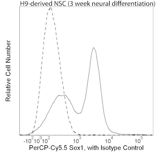
See MoreLEFT: Intracellular staining of Pax-6 in neural induction of human embryonic stem (ES) cells. H9 human ES cells (WiCell, Madison, WI) were cultured in mTeSR® (Stem Cell Technologies) on plates coated with BD Matrigel™ hESC-qualified Matrix (Cat. No. 354277). Embryoid bodies (EB) were made and cultured in medium containing Knockout™ Serum Replacement (Life Technologies) without bFGF for 24 hours and then in medium containing 250 ng/ml human recombinant noggin (R&D Systems) and 10 μM SB 431542 (Tocris) for 4 more days. The EB were then plated on BD Matrigel-coated plates and grown in medium with ITS supplement (Sigma-Aldrich), noggin, and SB 431542. After growth for 7 days, the cells were collected, fixed with BD Cytofix™ Fixation Buffer (Cat. No. 554655), and permeabilized with BD Phosflow™ Perm Buffer III (Cat. No. 558050). The cells were then stained with Alexa Fluor® 488 Mouse anti-Human Pax-6 (Cat. No. 561664) and PerCP-Cy™5.5 Mouse anti-Human Sox1 (Cat. No. 561549) antibodies. The plot was derived from gated events based on light scattering characteristics for the neural induction. Flow cytometry was performed on a BD™ LSR II flow cytometry system.
RIGHT: Immunofluorescent staining of Pax-6 in human embryonic stem (ES) cell-derived rosettes. H9 human ES cells (WiCell Madison, WI) grown on irradiated mouse embryonic fibroblasts were differentiated towards a neural stem cell lineage using a combination of 250 ng/ml human recombinant noggin (R&D systems) and 600 nM dorsomorphin (Sigma- Aldrich). The cells were fixed with BD CytofixT Fixation Buffer (Cat. No. 554655), permeabilized with 0.1% Triton™ X-100 Buffer, and stained with Alexa Fluor® 488 Mouse anti-Human Pax-6 (pseudo-colored green). Counter-staining was with Hoechst 33342 (pseudo-colored blue). The images were captured on a BD Pathway™ 435 Cell Analyzer and merged using BD Attovision™ Software.
LEFT: Intracellular staining of Pax-6 in neural induction of human embryonic stem (ES) cells. H9 human ES cells (WiCell, Madison, WI) were cultured in mTeSR® (Stem Cell Technologies) on plates coated with BD Matrigel™ hESC-qualified Matrix (Cat. No. 354277). Embryoid bodies (EB) were made and cultured in medium containing Knockout™ Serum Replacement (Life Technologies) without bFGF for 24 hours and then in medium containing 250 ng/ml human recombinant noggin (R&D Systems) and 10 μM SB 431542 (Tocris) for 4 more days. The EB were then plated on BD Matrigel-coated plates and grown in medium with ITS supplement (Sigma-Aldrich), noggin, and SB 431542. After growth for 7 days, the cells were collected, fixed with BD Cytofix™ Fixation Buffer (Cat. No. 554655), and permeabilized with BD Phosflow™ Perm Buffer III (Cat. No. 558050). The cells were then stained with Alexa Fluor® 488 Mouse anti-Human Pax-6 (Cat. No. 561664) and PerCP-Cy™5.5 Mouse anti-Human Sox1 (Cat. No. 561549) antibodies. The plot was derived from gated events based on light scattering characteristics for the neural induction. Flow cytometry was performed on a BD™ LSR II flow cytometry system.
RIGHT: Immunofluorescent staining of Pax-6 in human embryonic stem (ES) cell-derived rosettes. H9 human ES cells (WiCell Madison, WI) grown on irradiated mouse embryonic fibroblasts were differentiated towards a neural stem cell lineage using a combination of 250 ng/ml human recombinant noggin (R&D systems) and 600 nM dorsomorphin (Sigma- Aldrich). The cells were fixed with BD CytofixT Fixation Buffer (Cat. No. 554655), permeabilized with 0.1% Triton™ X-100 Buffer, and stained with Alexa Fluor® 488 Mouse anti-Human Pax-6 (pseudo-colored green). Counter-staining was with Hoechst 33342 (pseudo-colored blue). The images were captured on a BD Pathway™ 435 Cell Analyzer and merged using BD Attovision™ Software.
LEFT: Intracellular staining of Pax-6 in neural induction of human embryonic stem (ES) cells. H9 human ES cells (WiCell, Madison, WI) were cultured in mTeSR® (Stem Cell Technologies) on plates coated with BD Matrigel™ hESC-qualified Matrix (Cat. No. 354277). Embryoid bodies (EB) were made and cultured in medium containing Knockout™ Serum Replacement (Life Technologies) without bFGF for 24 hours and then in medium containing 250 ng/ml human recombinant noggin (R&D Systems) and 10 μM SB 431542 (Tocris) for 4 more days. The EB were then plated on BD Matrigel-coated plates and grown in medium with ITS supplement (Sigma-Aldrich), noggin, and SB 431542. After growth for 7 days, the cells were collected, fixed with BD Cytofix™ Fixation Buffer (Cat. No. 554655), and permeabilized with BD Phosflow™ Perm Buffer III (Cat. No. 558050). The cells were then stained with Alexa Fluor® 488 Mouse anti-Human Pax-6 (Cat. No. 561664) and PerCP-Cy™5.5 Mouse anti-Human Sox1 (Cat. No. 561549) antibodies. The plot was derived from gated events based on light scattering characteristics for the neural induction. Flow cytometry was performed on a BD™ LSR II flow cytometry system.
RIGHT: Immunofluorescent staining of Pax-6 in human embryonic stem (ES) cell-derived rosettes. H9 human ES cells (WiCell Madison, WI) grown on irradiated mouse embryonic fibroblasts were differentiated towards a neural stem cell lineage using a combination of 250 ng/ml human recombinant noggin (R&D systems) and 600 nM dorsomorphin (Sigma- Aldrich). The cells were fixed with BD CytofixT Fixation Buffer (Cat. No. 554655), permeabilized with 0.1% Triton™ X-100 Buffer, and stained with Alexa Fluor® 488 Mouse anti-Human Pax-6 (pseudo-colored green). Counter-staining was with Hoechst 33342 (pseudo-colored blue). The images were captured on a BD Pathway™ 435 Cell Analyzer and merged using BD Attovision™ Software.
LEFT: Intracellular staining of Pax-6 in neural induction of human embryonic stem (ES) cells. H9 human ES cells (WiCell, Madison, WI) were cultured in mTeSR® (Stem Cell Technologies) on plates coated with BD Matrigel™ hESC-qualified Matrix (Cat. No. 354277). Embryoid bodies (EB) were made and cultured in medium containing Knockout™ Serum Replacement (Life Technologies) without bFGF for 24 hours and then in medium containing 250 ng/ml human recombinant noggin (R&D Systems) and 10 μM SB 431542 (Tocris) for 4 more days. The EB were then plated on BD Matrigel-coated plates and grown in medium with ITS supplement (Sigma-Aldrich), noggin, and SB 431542. After growth for 7 days, the cells were collected, fixed with BD Cytofix™ Fixation Buffer (Cat. No. 554655), and permeabilized with BD Phosflow™ Perm Buffer III (Cat. No. 558050). The cells were then stained with Alexa Fluor® 488 Mouse anti-Human Pax-6 (Cat. No. 561664) and PerCP-Cy™5.5 Mouse anti-Human Sox1 (Cat. No. 561549) antibodies. The plot was derived from gated events based on light scattering characteristics for the neural induction. Flow cytometry was performed on a BD™ LSR II flow cytometry system.
RIGHT: Immunofluorescent staining of Pax-6 in human embryonic stem (ES) cell-derived rosettes. H9 human ES cells (WiCell Madison, WI) grown on irradiated mouse embryonic fibroblasts were differentiated towards a neural stem cell lineage using a combination of 250 ng/ml human recombinant noggin (R&D systems) and 600 nM dorsomorphin (Sigma- Aldrich). The cells were fixed with BD CytofixT Fixation Buffer (Cat. No. 554655), permeabilized with 0.1% Triton™ X-100 Buffer, and stained with Alexa Fluor® 488 Mouse anti-Human Pax-6 (pseudo-colored green). Counter-staining was with Hoechst 33342 (pseudo-colored blue). The images were captured on a BD Pathway™ 435 Cell Analyzer and merged using BD Attovision™ Software.
LEFT: Intracellular staining of Pax-6 in neural induction of human embryonic stem (ES) cells. H9 human ES cells (WiCell, Madison, WI) were cultured in mTeSR® (Stem Cell Technologies) on plates coated with BD Matrigel™ hESC-qualified Matrix (Cat. No. 354277). Embryoid bodies (EB) were made and cultured in medium containing Knockout™ Serum Replacement (Life Technologies) without bFGF for 24 hours and then in medium containing 250 ng/ml human recombinant noggin (R&D Systems) and 10 μM SB 431542 (Tocris) for 4 more days. The EB were then plated on BD Matrigel-coated plates and grown in medium with ITS supplement (Sigma-Aldrich), noggin, and SB 431542. After growth for 7 days, the cells were collected, fixed with BD Cytofix™ Fixation Buffer (Cat. No. 554655), and permeabilized with BD Phosflow™ Perm Buffer III (Cat. No. 558050). The cells were then stained with Alexa Fluor® 488 Mouse anti-Human Pax-6 (Cat. No. 561664) and PerCP-Cy™5.5 Mouse anti-Human Sox1 (Cat. No. 561549) antibodies. The plot was derived from gated events based on light scattering characteristics for the neural induction. Flow cytometry was performed on a BD™ LSR II flow cytometry system.
RIGHT: Immunofluorescent staining of Pax-6 in human embryonic stem (ES) cell-derived rosettes. H9 human ES cells (WiCell Madison, WI) grown on irradiated mouse embryonic fibroblasts were differentiated towards a neural stem cell lineage using a combination of 250 ng/ml human recombinant noggin (R&D systems) and 600 nM dorsomorphin (Sigma- Aldrich). The cells were fixed with BD CytofixT Fixation Buffer (Cat. No. 554655), permeabilized with 0.1% Triton™ X-100 Buffer, and stained with Alexa Fluor® 488 Mouse anti-Human Pax-6 (pseudo-colored green). Counter-staining was with Hoechst 33342 (pseudo-colored blue). The images were captured on a BD Pathway™ 435 Cell Analyzer and merged using BD Attovision™ Software.
LEFT: Intracellular staining of Pax-6 in neural induction of human embryonic stem (ES) cells. H9 human ES cells (WiCell, Madison, WI) were cultured in mTeSR® (Stem Cell Technologies) on plates coated with BD Matrigel™ hESC-qualified Matrix (Cat. No. 354277). Embryoid bodies (EB) were made and cultured in medium containing Knockout™ Serum Replacement (Life Technologies) without bFGF for 24 hours and then in medium containing 250 ng/ml human recombinant noggin (R&D Systems) and 10 μM SB 431542 (Tocris) for 4 more days. The EB were then plated on BD Matrigel-coated plates and grown in medium with ITS supplement (Sigma-Aldrich), noggin, and SB 431542. After growth for 7 days, the cells were collected, fixed with BD Cytofix™ Fixation Buffer (Cat. No. 554655), and permeabilized with BD Phosflow™ Perm Buffer III (Cat. No. 558050). The cells were then stained with Alexa Fluor® 488 Mouse anti-Human Pax-6 (Cat. No. 561664) and PerCP-Cy™5.5 Mouse anti-Human Sox1 (Cat. No. 561549) antibodies. The plot was derived from gated events based on light scattering characteristics for the neural induction. Flow cytometry was performed on a BD™ LSR II flow cytometry system.
RIGHT: Immunofluorescent staining of Pax-6 in human embryonic stem (ES) cell-derived rosettes. H9 human ES cells (WiCell Madison, WI) grown on irradiated mouse embryonic fibroblasts were differentiated towards a neural stem cell lineage using a combination of 250 ng/ml human recombinant noggin (R&D systems) and 600 nM dorsomorphin (Sigma- Aldrich). The cells were fixed with BD CytofixT Fixation Buffer (Cat. No. 554655), permeabilized with 0.1% Triton™ X-100 Buffer, and stained with Alexa Fluor® 488 Mouse anti-Human Pax-6 (pseudo-colored green). Counter-staining was with Hoechst 33342 (pseudo-colored blue). The images were captured on a BD Pathway™ 435 Cell Analyzer and merged using BD Attovision™ Software.
LEFT: Intracellular staining of Pax-6 in neural induction of human embryonic stem (ES) cells. H9 human ES cells (WiCell, Madison, WI) were cultured in mTeSR® (Stem Cell Technologies) on plates coated with BD Matrigel™ hESC-qualified Matrix (Cat. No. 354277). Embryoid bodies (EB) were made and cultured in medium containing Knockout™ Serum Replacement (Life Technologies) without bFGF for 24 hours and then in medium containing 250 ng/ml human recombinant noggin (R&D Systems) and 10 μM SB 431542 (Tocris) for 4 more days. The EB were then plated on BD Matrigel-coated plates and grown in medium with ITS supplement (Sigma-Aldrich), noggin, and SB 431542. After growth for 7 days, the cells were collected, fixed with BD Cytofix™ Fixation Buffer (Cat. No. 554655), and permeabilized with BD Phosflow™ Perm Buffer III (Cat. No. 558050). The cells were then stained with Alexa Fluor® 488 Mouse anti-Human Pax-6 (Cat. No. 561664) and PerCP-Cy™5.5 Mouse anti-Human Sox1 (Cat. No. 561549) antibodies. The plot was derived from gated events based on light scattering characteristics for the neural induction. Flow cytometry was performed on a BD™ LSR II flow cytometry system.
RIGHT: Immunofluorescent staining of Pax-6 in human embryonic stem (ES) cell-derived rosettes. H9 human ES cells (WiCell Madison, WI) grown on irradiated mouse embryonic fibroblasts were differentiated towards a neural stem cell lineage using a combination of 250 ng/ml human recombinant noggin (R&D systems) and 600 nM dorsomorphin (Sigma- Aldrich). The cells were fixed with BD CytofixT Fixation Buffer (Cat. No. 554655), permeabilized with 0.1% Triton™ X-100 Buffer, and stained with Alexa Fluor® 488 Mouse anti-Human Pax-6 (pseudo-colored green). Counter-staining was with Hoechst 33342 (pseudo-colored blue). The images were captured on a BD Pathway™ 435 Cell Analyzer and merged using BD Attovision™ Software.
LEFT: Intracellular staining of Pax-6 in neural induction of human embryonic stem (ES) cells. H9 human ES cells (WiCell, Madison, WI) were cultured in mTeSR® (Stem Cell Technologies) on plates coated with BD Matrigel™ hESC-qualified Matrix (Cat. No. 354277). Embryoid bodies (EB) were made and cultured in medium containing Knockout™ Serum Replacement (Life Technologies) without bFGF for 24 hours and then in medium containing 250 ng/ml human recombinant noggin (R&D Systems) and 10 μM SB 431542 (Tocris) for 4 more days. The EB were then plated on BD Matrigel-coated plates and grown in medium with ITS supplement (Sigma-Aldrich), noggin, and SB 431542. After growth for 7 days, the cells were collected, fixed with BD Cytofix™ Fixation Buffer (Cat. No. 554655), and permeabilized with BD Phosflow™ Perm Buffer III (Cat. No. 558050). The cells were then stained with Alexa Fluor® 488 Mouse anti-Human Pax-6 (Cat. No. 561664) and PerCP-Cy™5.5 Mouse anti-Human Sox1 (Cat. No. 561549) antibodies. The plot was derived from gated events based on light scattering characteristics for the neural induction. Flow cytometry was performed on a BD™ LSR II flow cytometry system.
RIGHT: Immunofluorescent staining of Pax-6 in human embryonic stem (ES) cell-derived rosettes. H9 human ES cells (WiCell Madison, WI) grown on irradiated mouse embryonic fibroblasts were differentiated towards a neural stem cell lineage using a combination of 250 ng/ml human recombinant noggin (R&D systems) and 600 nM dorsomorphin (Sigma- Aldrich). The cells were fixed with BD CytofixT Fixation Buffer (Cat. No. 554655), permeabilized with 0.1% Triton™ X-100 Buffer, and stained with Alexa Fluor® 488 Mouse anti-Human Pax-6 (pseudo-colored green). Counter-staining was with Hoechst 33342 (pseudo-colored blue). The images were captured on a BD Pathway™ 435 Cell Analyzer and merged using BD Attovision™ Software.
For Bioimaging procedures, please refer to the protocols under "Cellular Imaging" at our website: http://www.bdbiosciences.com/us/s/resources
Pax-6 is a member of the paired box (pax) gene family whose protein products are transcription factors involved in development. Pax family members share a highly conserved DNA binding domain that contains six alpha helices (paired domain) and a homeo box domain. Pax-6 has important roles in the development of the eye, nose, central nervous system, and pancreas. Defects in Pax-6 are responsible for various eye malformations including aniridia and Peters anomaly.
The O18-1330 monoclonal antibody reacts with human Pax-6. Because the Pax-6 protein sequence is highly conserved among vertebrate species, cross-reactivity with other species is possible.
Please refer to Support Documents for Quality Certificates
Global - Refer to manufacturer's instructions for use and related User Manuals and Technical data sheets before using this products as described
Comparisons, where applicable, are made against older BD Technology, manual methods or are general performance claims. Comparisons are not made against non-BD technologies, unless otherwise noted.
Please refer to Support Documents for Quality Certificates
Global - Refer to manufacturer's instructions for use and related User Manuals and Technical data sheets before using this products as described
Comparisons, where applicable, are made against older BD Technology, manual methods or are general performance claims. Comparisons are not made against non-BD technologies, unless otherwise noted.
For Research Use Only. Not for use in diagnostic or therapeutic procedures.