BD Horizon™ R718 Rat Anti-Mouse CD71
Clone RI7217 (also known as RI7 217.1.3; R17 217.1.4; R17 217; R17217)
(RUO)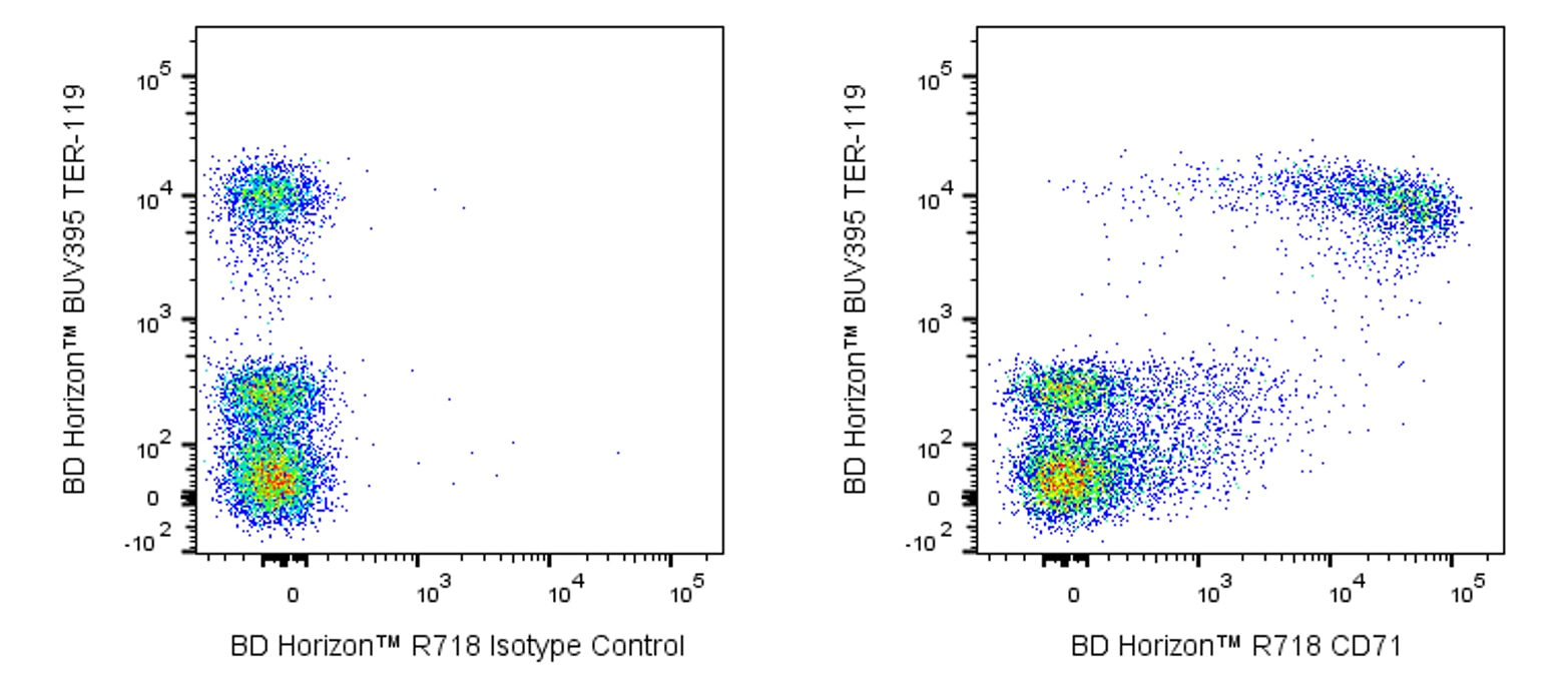
Preparation And Storage
Recommended Assay Procedures
BD™ CompBeads can be used as surrogates to assess fluorescence spillover (Compensation). When fluorochrome conjugated antibodies are bound to BD CompBeads, they have spectral properties very similar to cells. However, for some fluorochromes there can be small differences in spectral emissions compared to cells, resulting in spillover values that differ when compared to biological controls. It is strongly recommended that when using a reagent for the first time, users compare the spillover on cells and BD CompBead to ensure that BD CompBeads are appropriate for your specific cellular application.
Product Notices
- Since applications vary, each investigator should titrate the reagent to obtain optimal results.
- An isotype control should be used at the same concentration as the antibody of interest.
- Caution: Sodium azide yields highly toxic hydrazoic acid under acidic conditions. Dilute azide compounds in running water before discarding to avoid accumulation of potentially explosive deposits in plumbing.
- This product is provided under an Agreement between BIOTIUM and BD Biosciences. This product, and only in the amount purchased by buyer, may be used solely for buyer’s own internal research, in a manner consistent with the accompanying product literature. No other right to use, sell or otherwise transfer (a) this product, or (b) its components is hereby granted expressly, by implication or by estoppel. This product is for research use only. Diagnostic uses require a separate license from Biotium, Inc. For information on purchasing a license to this product including for purposes other than research, contact Biotium, Inc., 3159 Corporate Place, Hayward, CA 94545, Tel: (510) 265-1027. Fax: (510) 265-1352. Email: btinfo@biotium.com.
- Please refer to http://regdocs.bd.com to access safety data sheets (SDS).
- Alexa Fluor™ is a trademark of Life Technologies Corporation.
- Please refer to www.bdbiosciences.com/us/s/resources for technical protocols.
Companion Products


.png?imwidth=320)
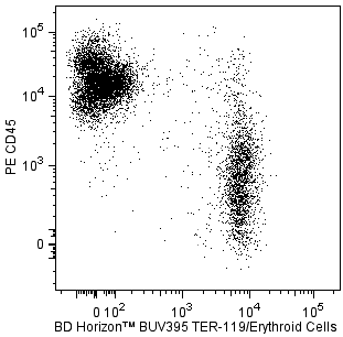
The RI7217 monoclonal antibody specifically recognizes the mouse Transferrin Receptor (TfR, Trfr, or TR), which is also known as Transferrin Receptor protein 1 (TfR1), CD71, and Mammary tumor virus receptor 1 (Mtvr-1 or Mtvr1). CD71 is a ~95-kDa single-pass type II transmembrane glycoprotein that is encoded by Tfrc and expressed on the cell surface as a disulfide-linked homodimer. The CD71 monomer is comprised of an extracellular C-terminal domain with a transferrin binding site followed by a transmembrane region and an N-terminal cytoplasmic tail that is involved in mediating the endocytosis and recycling of the receptor. This receptor mediates cellular iron uptake via endocytosis of an iron-transferrin complex followed by the recycling of transferrin and the receptor to the cell surface. CD71 is lowly expressed on most nonactivated cells including resting B and T lymphocytes. CD71 expression is upregulated on activated and proliferating cells such as mitogen- or antigen-stimulated B cells and T cells as well as some tumor cells. CD71 is variably expressed on erythroid precursors from pronormoblasts (proerythroblasts) to reticulocytes and is absent on mature red blood cells. CD71 is expressed on erythroid precursors that utilize iron for heme synthesis. CD71 can also serve as an entry receptor for several viral infections including those caused by Mouse mammary tumor virus (MMTV). The RI7217 antibody can reportedly be used to inhibit cellular proliferation.
The antibody was conjugated to BD Horizon Red 718, which has been developed exclusively by for BD Biosciences as a better alternative to Alexa Fluor™ 700. BD Horizon Red 718 can be excited by the red laser (628 – 640 nm) and, with an Em Max around 718 nm, it can be detected using a 730/45 nm filter. Due to similar excitation and emission properties, we do not recommend using R718 in combination with APC-R700 or Alexa Fluor™ 700.

Development References (6)
-
Ernst DN, McQuitty DN, Weigle WO, Hobbs MV. Expression of membrane activation antigens on murine B lymphocytes stimulated with lipopolysaccharide. Cell Immunol. 1988; 114(1):161-173. (Clone-specific: Flow cytometry). View Reference
-
Ernst DN, Weigle WO, McQuitty DN, Rothermel AL, Hobbs MV. Stimulation of murine T cell subsets with anti-CD3 antibody. Age-related defects in the expression of early activation molecules. J Immunol. 1989; 142(5):1413-1421. (Clone-specific: Flow cytometry). View Reference
-
Kemp JD, Thorson JA, Gomez F, Smith KM, Cowdery JS, Ballas ZK. Inhibition of lymphocyte activation with anti-transferrin receptor Mabs: a comparison of three reagents and further studies of their range of effects and mechanism of action. Cell Immunol. 1989; 122(1):218-230. (Clone-specific: Functional assay, Inhibition). View Reference
-
Lesley J, Hyman R, Schulte R, Trotter J. Expression of transferrin receptor on murine hematopoietic progenitors.. Cell Immunol. 1984; 83(1):14-25. (Immunogen: Flow cytometry). View Reference
-
Lesley JF, Schulte RJ. Inhibition of cell growth by monoclonal anti-transferrin receptor antibodies.. Mol Cell Biol. 1985; 5(8):1814-21. (Clone-specific: Blocking, Flow cytometry, Functional assay, Inhibition). View Reference
-
Motamedi M, Xu L, Elahi S. Correlation of transferrin receptor (CD71) with Ki67 expression on stimulated human and mouse T cells: The kinetics of expression of T cell activation markers.. J Immunol Methods. 2016; 437:43-52. (Biology: Flow cytometry). View Reference
Please refer to Support Documents for Quality Certificates
Global - Refer to manufacturer's instructions for use and related User Manuals and Technical data sheets before using this products as described
Comparisons, where applicable, are made against older BD Technology, manual methods or are general performance claims. Comparisons are not made against non-BD technologies, unless otherwise noted.
For Research Use Only. Not for use in diagnostic or therapeutic procedures.
Refer to manufacturer's instructions for use and related User Manuals and Technical Data Sheets before using this product as described.
Comparisons, where applicable, are made against older BD technology, manual methods or are general performance claims. Comparisons are not made against non-BD technologies, unless otherwise noted.