Skip to main content Skip to navigation
PE Mouse Anti-MR1
PE Mouse Anti-MR1
Flow cytometric analysis of MR1 expression on human peripheral blood lymphocytes. Human peripheral blood mononuclear cells (PBMCs) were either cultured (16 h) with Acetyl-6-Formylpterin (+ Ac-6-FP; 2.3 µg/ml (10 µM); Cayman Chemicals Cat. No. 23303) or without Ac-6-FP (No Ac-6-FP) as indicated. The cells were then preincubated with Human BD Fc Block™ (Cat. No. 564219/564220) and stained with either PE Mouse IgG1, κ Isotype Control (Cat. No. 554680; dotted line histograms) or PE Mouse anti-MR1 antibody (Cat. No. 568012/568013; solid line histograms) at 0.5 µg/test. DAPI Solution (Cat. No. 564907) was added to cells right before analysis. The fluorescence histograms showing MR1 expression (or Ig Isotype control staining) were derived from gated events with the forward and side light-scatter characteristics of viable (DAPI-negative) lymphocytes. Flow cytometry and data analysis were performed using a BD LSRFortessa™ X-20 Cell Analyzer System and FlowJo™ software. Data shown on this Technical Data Sheet are not lot specific.
PE Mouse Anti-MR1
Flow cytometric analysis of MR1 expression on B16-F0 cells. Cells from the mouse B16-F0 (Skin Melanoma, ATCC CRL-6322) cell line were either cultured (16 h) with Acetyl-6-Formylpterin (+ Ac-6-FP; 2.3 µg/ml (10 µM); Cayman Chemicals Cat. No. 23303) or without Ac-6-FP (No Ac-6-FP) as indicated. The cells were then preincubated with Purified Rat Anti-Mouse CD16/CD32 antibody (Mouse BD Fc Block™) (Cat. No. 553141/553142) and stained with either PE Mouse IgG1, κ Isotype Control (Cat. No. 554680; dotted line histograms) or PE Mouse Anti-MR1 antibody (Cat. No. 568012/568013; solid line histograms) at 0.5 µg/test. DAPI Solution (Cat. No. 564907) was added to cells right before analysis. The fluorescence histograms showing MR1 expression (or Ig Isotype control staining) were derived from gated events with the forward and side light-scatter characteristics of viable (DAPI-negative) cells. Flow cytometry and data analysis were performed using a BD LSRFortessa™ X20 Cell Analyzer System and FlowJo™ software. Data shown on this Technical Data Sheet are not lot specific.
Flow cytometric analysis of MR1 expression on human peripheral blood lymphocytes. Human peripheral blood mononuclear cells (PBMCs) were either cultured (16 h) with Acetyl-6-Formylpterin (+ Ac-6-FP; 2.3 µg/ml (10 µM); Cayman Chemicals Cat. No. 23303) or without Ac-6-FP (No Ac-6-FP) as indicated. The cells were then preincubated with Human BD Fc Block™ (Cat. No. 564219/564220) and stained with either PE Mouse IgG1, κ Isotype Control (Cat. No. 554680; dotted line histograms) or PE Mouse anti-MR1 antibody (Cat. No. 568012/568013; solid line histograms) at 0.5 µg/test. DAPI Solution (Cat. No. 564907) was added to cells right before analysis. The fluorescence histograms showing MR1 expression (or Ig Isotype control staining) were derived from gated events with the forward and side light-scatter characteristics of viable (DAPI-negative) lymphocytes. Flow cytometry and data analysis were performed using a BD LSRFortessa™ X-20 Cell Analyzer System and FlowJo™ software. Data shown on this Technical Data Sheet are not lot specific.
Flow cytometric analysis of MR1 expression on B16-F0 cells. Cells from the mouse B16-F0 (Skin Melanoma, ATCC CRL-6322) cell line were either cultured (16 h) with Acetyl-6-Formylpterin (+ Ac-6-FP; 2.3 µg/ml (10 µM); Cayman Chemicals Cat. No. 23303) or without Ac-6-FP (No Ac-6-FP) as indicated. The cells were then preincubated with Purified Rat Anti-Mouse CD16/CD32 antibody (Mouse BD Fc Block™) (Cat. No. 553141/553142) and stained with either PE Mouse IgG1, κ Isotype Control (Cat. No. 554680; dotted line histograms) or PE Mouse Anti-MR1 antibody (Cat. No. 568012/568013; solid line histograms) at 0.5 µg/test. DAPI Solution (Cat. No. 564907) was added to cells right before analysis. The fluorescence histograms showing MR1 expression (or Ig Isotype control staining) were derived from gated events with the forward and side light-scatter characteristics of viable (DAPI-negative) cells. Flow cytometry and data analysis were performed using a BD LSRFortessa™ X20 Cell Analyzer System and FlowJo™ software. Data shown on this Technical Data Sheet are not lot specific.
Product Details
Down Arrow Up Arrow


BD Pharmingen™
HLALS; MHC class I-like antigen MR-1; MHC class-I related-gene protein
Human (QC Testing), Mouse (Tested in Development)
Mouse IgG1, κ
MR1 Extracellular Domain Complexed with β2m
Flow cytometry (Routinely Tested)
0.2 mg/ml
Aqueous buffered solution containing ≤0.09% sodium azide.
RUO


Preparation And Storage

Store undiluted at 4°C and protected from prolonged exposure to light. Do not freeze. The monoclonal antibody was purified from tissue culture supernatant or ascites by affinity chromatography. The antibody was conjugated to the dye under optimum conditions and unconjugated antibody and free dye were removed.

Recommended Assay Procedures

BD® CompBeads can be used as surrogates to assess fluorescence spillover (Compensation). When fluorochrome conjugated antibodies are bound to BD® CompBeads, they have spectral properties very similar to cells. However, for some fluorochromes there can be small differences in spectral emissions compared to cells, resulting in spillover values that differ when compared to biological controls. It is strongly recommended that when using a reagent for the first time, users compare the spillover on cells and BD CompBeads to ensure that BD® CompBeads are appropriate for your specific cellular application.

Product Notices

  1. Please refer to www.bdbiosciences.com/us/s/resources for technical protocols.
  2. Caution: Sodium azide yields highly toxic hydrazoic acid under acidic conditions. Dilute azide compounds in running water before discarding to avoid accumulation of potentially explosive deposits in plumbing.
  3. For fluorochrome spectra and suitable instrument settings, please refer to our Multicolor Flow Cytometry web page at www.bdbiosciences.com/colors.
  4. Please refer to http://regdocs.bd.com to access safety data sheets (SDS).
  5. Since applications vary, each investigator should titrate the reagent to obtain optimal results.
  6. An isotype control should be used at the same concentration as the antibody of interest.
Antibody Details
Down Arrow Up Arrow
8F2.F9

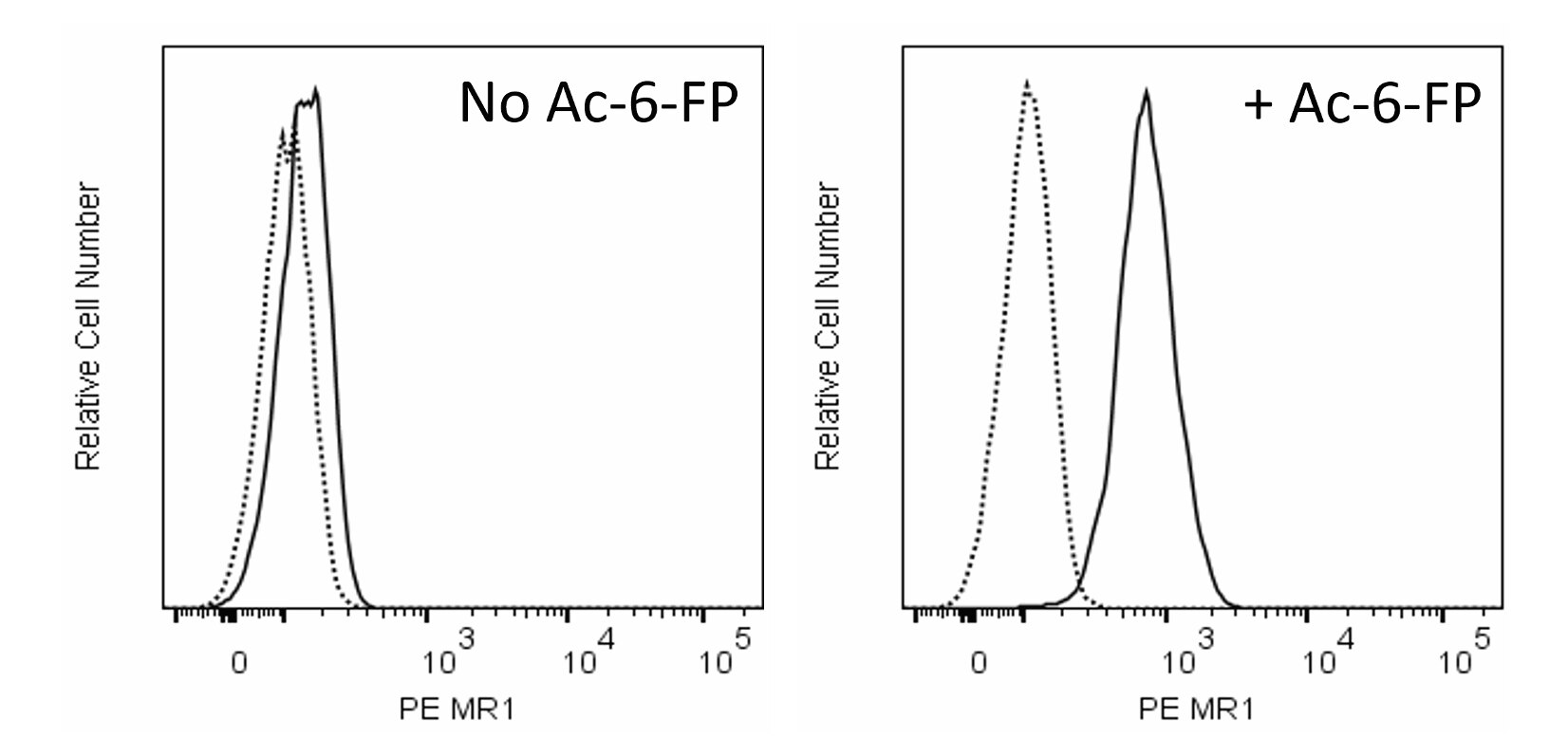
The 8F2.F9 monoclonal antibody specifically recognizes MHC-related protein 1 (MR1). MRI is a nonclassical MHC class Ib molecule. It is comprised of a ~40 kDa, highly conserved transmembrane a heavy chain that is a type I glycoprotein which is noncovalently-associated with an invariant ß2-microglobulin (ß2m) light chain. The N-terminal extracellular region of the HLA class I heavy chain is comprised of three domains (a1, a2, and a3). The a1 and a2 domains form a closed antigen-binding groove that accommodates small antigens whereas the a3 domain interacts with ß2m. MR1 is an antigen-presenting molecule that is specialized in displaying metabolites derived from microbial riboflavin biosynthesis to a small population of aß T-cells expressing an invariant TCR a chain called mucosal-associated invariant T-cells (MAIT). This function is essential to the development and expansion of MAIT cells. The 8F2.F9 and 26.5 monoclonal antibodies reportedly bind to different MRI epitopes.

Format Details
Down Arrow Up Arrow
PE
R-Phycoerythrin (PE), is part of the BD family of Phycobiliprotein dyes. This fluorochrome is a multimeric fluorescent phycobiliprotein with excitation maximum (Ex Max) of 496 nm and 566 nm and an emission maximum (Em Max) at 576 nm. PE is designed to be excited by the Blue (488 nm), Green (532 nm) and Yellow-Green (561 nm) lasers and detected using an optical filter centered near 575 nm (e.g., a 575/26-nm bandpass filter). As PE is excited by multiple lasers, this can result in cross-laser excitation and fluorescence spillover on instruments with various combinations of Blue, Green, and Yellow-Green lasers. Please ensure that your instrument’s configurations (lasers and optical filters) are appropriate for this dye.
altImg
PE
Yellow-Green 488 nm, 532 nm, 561 nm
496 nm, 566 nm
576 nm
Citations & References
Down Arrow Up Arrow

Development References (9)

  1. Abós B, Gómez Del Moral M, Gozalbo-López B, López-Relaño J, Viana V, Martínez-Naves E. Human MR1 expression on the cell surface is acid sensitive, proteasome independent and increases after culturing at 26°C. Biochem Biophys Res Commun. 2011; 411(3):632-636. (Biology: Flow cytometry). View Reference
  2. Chua WJ, Kim S, Myers N, et al. Endogenous MHC-related protein 1 is transiently expressed on the plasma membrane in a conformation that activates mucosal-associated invariant T cells. J Immunol. 2011; 186(8):4744-4750. (Immunogen). View Reference
  3. Corbett AJ, Awad W, Wang H, Chen Z. Antigen Recognition by MR1-Reactive T Cells; MAIT Cells, Metabolites, and Remaining Mysteries. Front Immunol. 2020; 11(1961):1-18. (Biology). View Reference
  4. Huang S, Gilfillan S, Cella M, et al. Evidence for MR1 antigen presentation to mucosal-associated invariant T cells. J Biol Chem. 2005; 280(22):21183-21193. (Immunogen: Flow cytometry). View Reference
  5. Lamichhane R, Ussher JE. Expression and trafficking of MR1. Immunology. 2017; 151(3):270-279. (Biology). View Reference
  6. McWilliam HE, Eckle SB, Theodossis A, et al. The intracellular pathway for the presentation of vitamin B-related antigens by the antigen-presenting molecule MR1. Nat Immunol. 2016; 17(5):531-537. (Clone-specific: Flow cytometry). View Reference
  7. Miley MJ, Truscott SM, Yu YY, et al. Biochemical features of the MHC-related protein 1 consistent with an immunological function. J Immunol. 2003; 170(12):6090-6098. (Biology: Blocking, Flow cytometry, In vivo exacerbation). View Reference
  8. Salio M, Awad W, Veerapen N, et al. Ligand-dependent downregulation of MR1 cell surface expression. Proc Natl Acad Sci U S A. 2020; 117(19):10465-10475. (Clone-specific: Flow cytometry). View Reference
  9. Yan J, Allen S, McDonald E, et al. MAIT Cells Promote Tumor Initiation, Growth, and Metastases via Tumor MR1. Cancer Discovery. 2020; 10(1):124-141. (Clone-specific). View Reference
View All (9) View Less

Please refer to Support Documents for Quality Certificates


Global - Refer to manufacturer's instructions for use and related User Manuals and Technical data sheets before using this products as described


Comparisons, where applicable, are made against older BD Technology, manual methods or are general performance claims.  Comparisons are not made against non-BD technologies, unless otherwise noted.

For Research Use Only. Not for use in diagnostic or therapeutic procedures.

Refer to manufacturer's instructions for use and related User Manuals and Technical Data Sheets before using this product as described.

Comparisons, where applicable, are made against older BD technology, manual methods or are general performance claims. Comparisons are not made against non-BD technologies, unless otherwise noted.